我们检测到您正在 美国 访问我们的网站。
我们针对您的国家(美国)提供单独版本的网站。

切换至 美国
 技术

容积二氧化碳图。 精密的二氧化碳监测

图:放大镜

有关更多见解。 容积 CO2 监测

容积二氧化碳图的时相、形状和曲线形态和计算衍生的测量可以告诉您下列重要信息:

  • 通气-灌注效率
  • 生理死腔比
  • 病人的代谢率 (Jaffe MB.Using the features of the time and volumetric capnogram for classification and prediction.J Clin Monit Comput.2017;31(1):19-41. doi:10.1007/s10877-016-9830-z1​)
CAPNOSTAT-5 主流式 CO2 传感器

一个强大的工具。 CO2 传感器

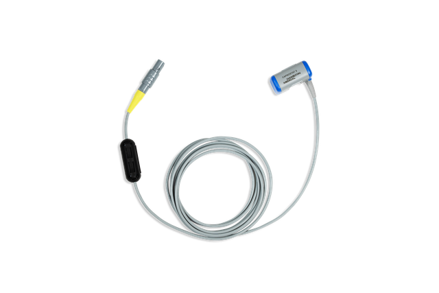
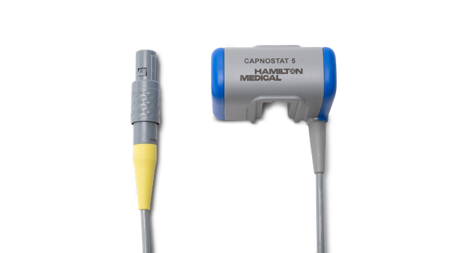
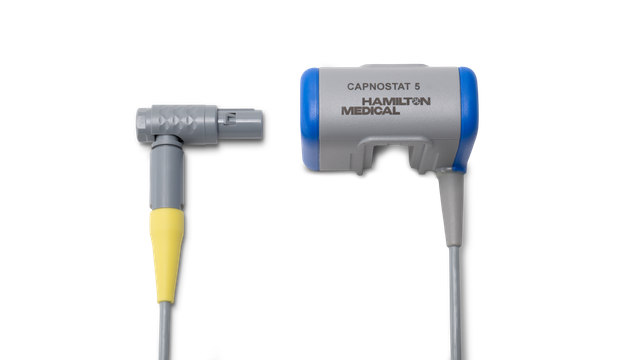
在我们的呼吸机上,使用病人气道近端的 CAPNOSTAT-5 主流式 CO2 传感器测量 CO2。

CAPNOSTAT-5 传感器针对呼气末二氧化碳分压 (PetCO2) 以及在 150 次/分钟下所有呼吸频率的清晰、准确的二氧化碳图提供准确的测量。

统计图:二氧化碳传感器数据分析

小传感器,大数据。 这就是您得到的数据

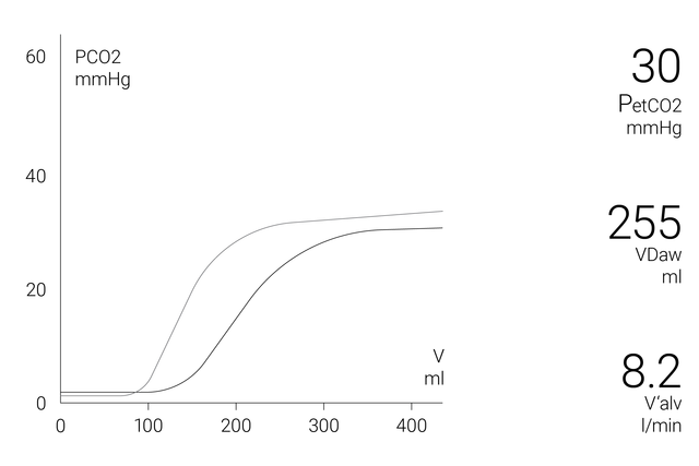
显示屏上的容积二氧化碳图窗口显示准确的定量信息作为近端流量和近端 CO2 数据的结合,例如:

  • 当前容积二氧化碳图曲线
  • 容积二氧化碳图参考曲线
  • 带有参考环时间和日期的参考曲线按钮
  • 每次呼吸的最相关 CO2 值

为了解更多关于病人状况的综合分析,可获得以下参数的 72 小时趋势图(或 HAMILTON-S1/G5 呼吸机的 96 小时趋势图):

  • PetCO2
  • V‘CO2
  • FetCO2
  • VeCO2
  • ViCO2
  • Vtalv
  • V'alv
  • VDaw
  • VD/Vt
  • VDaw/VTE
  • Slope CO2

为了使您一目了然,Hamilton Medical 哈美顿医疗公司呼吸机在二氧化碳监测窗口提供了所有 CO2 相关性数据的概览。

  • 呼气末二氧化碳浓度: FetCO2 (%) 
  • 呼气末二氧化碳压力:PetCO2 (mmHg) 
  • 在“PetCO2”曲线中的肺泡平台的斜率,表示肺的容量/流量状态: slopeCO2 (%CO2/l)
  • 肺泡潮气量:Vtalv (ml) 
  • 肺泡分钟通气量:V’alv (l/min) 
  • CO2清除状态:V’CO2 (ml/min) 
  • 气道死腔:VDaw (ml)
  • 气道开口处的气道死腔比:VDaw/VTE (%) 
  • 呼出的二氧化碳容量: VeCO2 (ml) 
  • 吸入的二氧化碳容量:ViCO2 (ml)
容积二氧化碳图电子书

免费电子书

不可不知! 容积二氧化碳图的详情

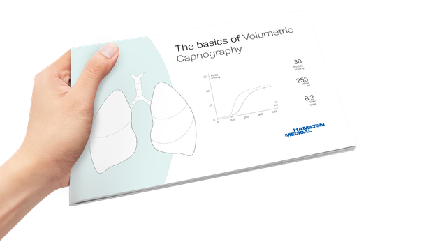
学习如何解读容积二氧化碳图及了解容积二氧化碳图的优点和临床应用。包括自测内容!

统计图:www.hamilton-medical.com/capnography

有哪些获益? 了解证据

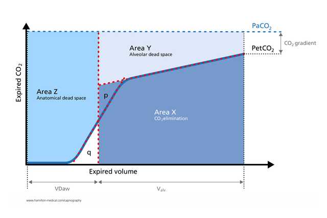
  • 容积二氧化碳图已成功用于测量解剖死腔、肺毛细血管灌注和通气效率 (Romero PV, Lucangelo U, Lopez Aguilar J, Fernandez R, Blanch L. Physiologically based indices of volumetric capnography in patients receiving mechanical ventilation.Eur Respir J. 1997;10(6):1309-1315. doi:10.1183/09031936.97.100613092​)

  • 容积二氧化碳图衍生计算有助于在床头识别肺动脉栓塞 (Blanch L, Romero PV, Lucangelo U. Volumetric capnography in the mechanically ventilated patient.Minerva Anestesiol.2006;72(6):577-585.3​)

  • 接受机械通气的 ARDS 病人生理死腔-潮气量比的容积二氧化碳图测量与通过代谢监测技术获得的数据一样准确 (Kallet RH, Daniel BM, Garcia O, Matthay MA.Accuracy of physiologic dead space measurements in patients with acute respiratory distress syndrome using volumetric capnography: comparison with the metabolic monitor method.Respir Care.2005;50(4):462-467.4​)

  • 呼气二氧化碳图是一种独立于呼吸努力、快速的无创测量,可帮助检测成人哮喘病人的显著支气管痉挛 (Yaron M, Padyk P, Hutsinpiller M, Cairns CB.Utility of the expiratory capnogram in the assessment of bronchospasm.Ann Emerg Med.1996;28(4):403-407. doi:10.1016/s0196-0644(96)70005-75​)

  • 容积二氧化碳图以无创、实时方式提供有关肺塌陷和肺复张生理学的宝贵见解,并有助于在床头监测周期性肺复张操作 (Tusman G, Suarez-Sipmann F, Böhm SH, et al.Monitoring dead space during recruitment and PEEP titration in an experimental model.Intensive Care Med.2006;32(11):1863-1871. doi:10.1007/s00134-006-0371-76​)

图:手持证书的学生

不可不知! 容积二氧化碳图培训资源

附件和耗材

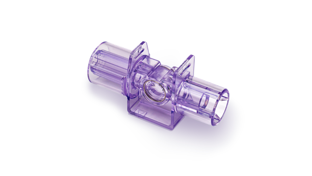
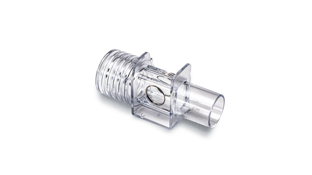
我们提供成人、儿童和新生儿病人用原装耗材。您可根据您的机构政策选择可重复使用和一次性使用产品。

可用性

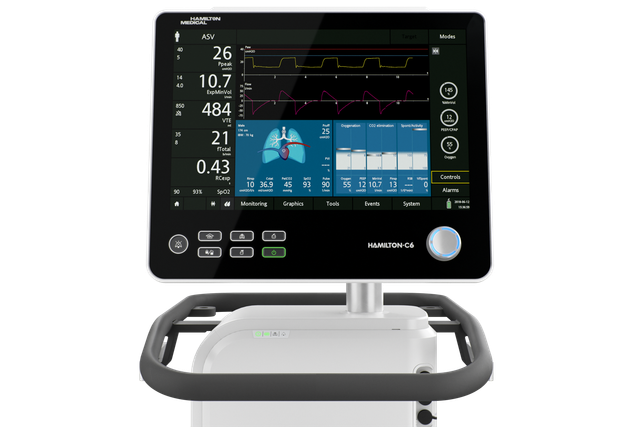
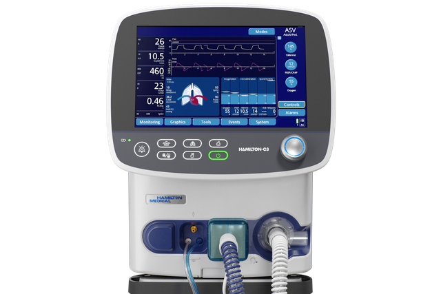
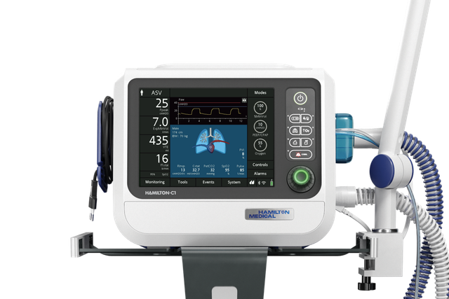
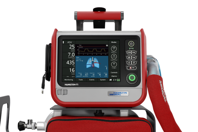
容积二氧化碳图可作为 HAMILTON-C6、HAMILTON-G5、HAMILTON-C3 和 HAMILTON-C1/T1 呼吸机的选配功能以及 HAMILTON-S1 呼吸机的标准功能提供。

Using the features of the time and volumetric capnogram for classification and prediction.

Jaffe MB. Using the features of the time and volumetric capnogram for classification and prediction. J Clin Monit Comput. 2017;31(1):19-41. doi:10.1007/s10877-016-9830-z

Quantitative features derived from the time-based and volumetric capnogram such as respiratory rate, end-tidal PCO2, dead space, carbon dioxide production, and qualitative features such as the shape of capnogram are clinical metrics recognized as important for assessing respiratory function. Researchers are increasingly exploring these and other known physiologically relevant quantitative features, as well as new features derived from the time and volumetric capnogram or transformations of these waveforms, for: (a) real-time waveform classification/anomaly detection, (b) classification of a candidate capnogram into one of several disease classes, (c) estimation of the value of an inaccessible or invasively determined physiologic parameter, (d) prediction of the presence or absence of disease condition, (e) guiding the administration of therapy, and (f) prediction of the likely future morbidity or mortality of a patient with a presenting condition. The work to date with respect to these applications will be reviewed, the underlying algorithms and performance highlighted, and opportunities for the future noted.

Physiologically based indices of volumetric capnography in patients receiving mechanical ventilation.

Romero PV, Lucangelo U, Lopez Aguilar J, Fernandez R, Blanch L. Physiologically based indices of volumetric capnography in patients receiving mechanical ventilation. Eur Respir J. 1997;10(6):1309-1315. doi:10.1183/09031936.97.10061309

Several indices of ventilatory heterogeneity can be identified from the expiratory CO2 partial pressure or CO2 elimination versus volume curves. The aims of this study were: 1) to analyse several computerizable indices of volumetric capnography in order to detect ventilatory disturbances; and 2) to establish the relationship between those indices and respiratory system mechanics in subjects with normal lungs and in patients with acute respiratory distress syndrome (ARDS), both receiving mechanical ventilation. We studied six normal subjects and five patients with early ARDS mechanically ventilated at three levels of tidal volume (VT). Respiratory system mechanics were assessed by end-expiratory and end-inspiratory occlusion methods, respectively. We determined Phase III slopes, Fletcher's efficiency index, Bohr's dead space (VD,Bohr/VT), and the ratio of alveolar ejection volume to tidal volume (VAE/VT) from expiratory capnograms, as a function of expired volume. Differences between normal subjects and ARDS patients were significant both for capnographic and mechanical parameters. Changes in VT significantly altered capnographic indices in normal subjects, but failed to change ventilatory mechanics and VAE/VT in ARDS patients. After adjusting for breathing pattern, VAE/VT exhibited the best correlation with the mechanical parameters. In conclusion, volumetric capnography, and, specifically, the ratio of alveolar ejection volume to tidal volume allows evaluation and monitoring of ventilatory disturbances in patients with adult respiratory distress syndrome.

Volumetric capnography in the mechanically ventilated patient.

Blanch L, Romero PV, Lucangelo U. Volumetric capnography in the mechanically ventilated patient. Minerva Anestesiol. 2006;72(6):577-585.

Expiratory capnogram provides qualitative information on the waveform patterns associated with mechanical ventilation and quantitative estimation of expired CO2. Volumetric capnography simultaneously measures expired CO2 and tidal volume and allows identification of CO2 from 3 sequential lung compartments: apparatus and anatomic dead space, from progressive emptying of alveoli and alveolar gas. Lung heterogeneity creates regional differences in CO2 concentration and sequential emptying contributes to the rise of the alveolar plateau and to the steeper the expired CO2 slope. The concept of dead space accounts for those lung areas that are ventilated but not perfused. In patients with sudden pulmonary vascular occlusion due to pulmonary embolism, the resultant high V/Q mismatch produces an increase in alveolar dead space. Calculations derived from volumetric capnography are useful to suspect pulmonary embolism at the bedside. Alveolar dead space is large in acute lung injury and when the effect of positive end-expiratory pressure (PEEP) is to recruit collapsed lung units resulting in an improvement of oxygenation, alveolar dead space may decrease, whereas PEEP-induced overdistension tends to increase alveolar dead space. Finally, measurement of physiologic dead space and alveolar ejection volume at admission or the trend during the first 48 hours of mechanical ventilation might provide useful information on outcome of critically ill patients with acute lung injury or acute respiratory distress syndrome.

Accuracy of physiologic dead space measurements in patients with acute respiratory distress syndrome using volumetric capnography: comparison with the metabolic monitor method.

Kallet RH, Daniel BM, Garcia O, Matthay MA. Accuracy of physiologic dead space measurements in patients with acute respiratory distress syndrome using volumetric capnography: comparison with the metabolic monitor method. Respir Care. 2005;50(4):462-467.


BACKGROUND

Volumetric capnography is an alternative method of measuring expired carbon dioxide partial pressure (P(eCO2)) and physiologic dead-space-to-tidal-volume ratio (V(D)/V(T)) during mechanical ventilation. In this method, P(eCO2) is measured at the Y-adapter of the ventilator circuit, thus eliminating the effects of compression volume contamination and the need to apply a correction factor. We investigated the accuracy of volumetric capnography in measuring V(D)/V(T), compared to both uncorrected and corrected measurements, using a metabolic monitor in patients with acute respiratory distress syndrome (ARDS).

METHODS

There were 90 measurements of V(D)/V(T) made in 23 patients with ARDS. The P(eCO2) was measured during a 5-min expired-gas collection period with a Delta-trac metabolic monitor, and was corrected for compression volume contamination using a standard formula. Simultaneous measurements of P(eCO2) and V(D)/V(T) were obtained using volumetric capnography.

RESULTS

V(D)/V(T) measured by volumetric capnography was strongly correlated with both the uncorrected (r2 = 0.93, p < 0.0001) and corrected (r2 = 0.89, p < 0.0001) measurements of V(D)/V(T) made using the metabolic monitor technique. Measurements of V(D)/V(T) made with volumetric capnography had a bias of 0.02 and a precision of 0.05 when compared to the V(D)/V(T) corrected for estimated compression volume contamination.

CONCLUSION

Volumetric capnography measurements of V(D)/V(T) in mechanically-ventilated patients with ARDS are as accurate as those obtained by metabolic monitor technique. .

Utility of the expiratory capnogram in the assessment of bronchospasm.

Yaron M, Padyk P, Hutsinpiller M, Cairns CB. Utility of the expiratory capnogram in the assessment of bronchospasm. Ann Emerg Med. 1996;28(4):403-407. doi:10.1016/s0196-0644(96)70005-7


STUDY OBJECTIVE

To determine whether the plateau phase of the expiratory capnogram (dco2/dt) can detect bronchospasm in adult asthma patients in the emergency department and to assess the correlation between dco2/dt and the peak expiratory flow rate (PEFR) in spontaneously breathing patients with asthma and in normal, healthy volunteers.

METHODS

We carried out a prospective, blinded study in a university hospital ED. Twenty adults (12 women) with acute asthma and 28 normal adult volunteers (15 women) breathed through the sampling probe of an end-tidal CO2 monitor, and the expired CO2 waveform was recorded. The dco2/dt of the plateau (alveolar) phase for five consecutive regular expirations was measured and a mean value calculated for each patient. The best of three PEFRs was determined. The PEFR and dco2/dt were also recorded after treatment of the asthmatic patients with inhaled beta-agonists.

RESULTS

The mean +/- SD PEFR of the asthmatic subjects was 274 +/- 96 L/minute (57% of the predicted value), whereas that of the normal volunteers was 527 +/- 96 L/minute (103% of the predicted value) (P < .001). The mean dco2/dt of the asthmatic subjects (.26 +/- .06) was significantly steeper than that of the normal volunteers (.13 +/- .06) (P < .001). The dco2/dt was correlated with PEFR (r = .84, P < .001). In 18 asthmatic subjects the pretreatment and posttreatment percent predicted PEFRS were 58% +/- 17% and 74% +/- 17%, respectively (P < .001), whereas the dco2/dt values were .27 +/- .05 and .19 +/- .07, respectively (P < .005).

CONCLUSION

The dco2/dt is an effort-independent, rapid noninvasive measure that indicates significant bronchospasm in ED adult patients with asthma. The dco2/dt value is correlated with PEFR, an effort-dependent measure of airway obstruction. The change in dco2/dt with inhaled beta-agonists may be useful in monitoring the therapy of acute asthma.

Monitoring dead space during recruitment and PEEP titration in an experimental model.

Tusman G, Suarez-Sipmann F, Böhm SH, et al. Monitoring dead space during recruitment and PEEP titration in an experimental model. Intensive Care Med. 2006;32(11):1863-1871. doi:10.1007/s00134-006-0371-7


OBJECTIVE

To test the usefulness of dead space for determining open-lung PEEP, the lowest PEEP that prevents lung collapse after a lung recruitment maneuver.

DESIGN

Prospective animal study.

SETTING

Department of Clinical Physiology, University of Uppsala, Sweden.

SUBJECTS

Eight lung-lavaged pigs.

INTERVENTIONS

Animals were ventilated using constant flow mode with VT of 6ml/kg, respiratory rate of 30bpm, inspiratory-to-expiratory ratio of 1:2, and FiO(2) of 1. Baseline measurements were performed at 6cmH(2)O of PEEP. PEEP was increased in steps of 6cmH(2)O from 6 to 24cmH(2)O. Recruitment maneuver was achieved within 2min at pressure levels of 60/30cmH(2)O for Peak/PEEP. PEEP was decreased from 24 to 6cmH(2)O in steps of 2cmH(2)O and then to 0cmH(2)O. Each PEEP step was maintained for 10min.

MEASUREMENTS AND RESULTS

Alveolar dead space (VD(alv)), the ratio of alveolar dead space to alveolar tidal volume (VD(alv)/VT(alv)), and the arterial to end-tidal PCO(2) difference (Pa-ET: CO(2)) showed a good correlation with PaO(2), normally aerated areas, and non-aerated CT areas in all animals (minimum-maximum r(2)=0.83-0.99; p<0.01). Lung collapse (non-aerated tissue>5%) started at 12[Symbol: see text]cmH(2)O PEEP; hence, open-lung PEEP was established at 14cmH(2)O. The receiver operating characteristics curve demonstrated a high specificity and sensitivity of VD(alv) (0.89 and 0.90), VD(alv)/VT(alv) (0.82 and 1.00), and Pa-ET: CO(2) (0.93 and 0.95) for detecting lung collapse.

CONCLUSIONS

Monitoring of dead space was useful for detecting lung collapse and for establishing open-lung PEEP after a recruitment maneuver.