Atrás

La capnografía de forma de onda básica como herramienta de monitorización continua durante la ventilación mecánica

Artículo

Autor: Joe Hylton, MA, BSRT, RRT-ACCS/NPS, NRP, FAARC, FCCM, especialista en aplicaciones clínicas, Hamilton Medical Inc.

Fecha: 15.07.2021

La capnografía de forma de onda es un procedimiento habitual en la medicina de cuidados intensivos. Se trata de una herramienta de validación del control de la vía aérea ampliamente utilizada y con mucho predicamento en los entornos de sedación consciente, así como durante los traslados entre instalaciones de pacientes intubados que requieren ventilación mecánica.  La capnografía de forma de onda puede proporcionar información oportuna y muy valiosa a un profesional sanitario con la debida formación.

La capnografía de forma de onda básica como herramienta de monitorización continua durante la ventilación mecánica

Factores fisiológicos que afectan al dióxido de carbono al final del volumen tidal (PetCO2)

Hay infinidad de factores que pueden afectar a la cantidad de dióxido de carbono en el gas al final del volumen tidal (PetCO2). Respecto a la eliminación de CO2, existe un estrecho equilibrio continuado entre la producción de CO2 en los tejidos, su transporte por la sangre, la difusión hacia los alveolos y la eliminación por ventilación (Kremeier P, Böhm SH, Tusman G. Clinical use of volumetric capnography in mechanically ventilated patients. J Clin Monit Comput. 2020;34(1):7-16. doi:10.1007/s10877-019-00325-91). La capnografía proporciona una representación gráfica del CO2 espirado y se puede utilizar como una forma no invasiva de mostrar información en tiempo real sobre la cinética del CO2 en pacientes con ventilación mecánica.

Un aumento o disminución de la tasa metabólica del paciente conllevaría un cambio en la producción de CO2 y, por tanto, también en la eliminación de CO2. Si tanto la circulación como la ventilación son estables —estado que solo pueden alcanzar los pacientes con ventilación mecánica pasivos—, la monitorización del CO2 se puede usar como indicador de la producción de CO2. La fiebre, la septicemia, el dolor o las convulsiones son todos cuadros que pueden aumentar la actividad metabólica, lo que conlleva el correspondiente aumento de la producción de CO2 y, por tanto, un valor de PetCO2 mayor. Por el contrario, la actividad metabólica disminuye en pacientes con hipotermia, sedados o paralizados. Esto reduce la producción de CO2 y puede desembocar en una disminución del valor de PetCO2 si la ventilación minuto no aumenta al mismo tiempo (Gravenstein, J., Jaffe, M., & Paulus, D. (2004). Capnography: Clinical Aspects. New York: Cambridge University Press.2).

El transporte de CO2 a los pulmones depende de una correcta función cardiovascular. En consecuencia, cualquier factor que altere la función cardiovascular puede influir también en el transporte de CO2 a los pulmones (Gravenstein, J., Jaffe, M., & Paulus, D. (2004). Capnography: Clinical Aspects. New York: Cambridge University Press.2).

La expulsión del CO2 de los pulmones al entorno exterior se ve afectada por los cambios en la función respiratoria. Las enfermedades pulmonares obstructivas, la neumonía, los trastornos neuromusculares y los trastornos del sistema nervioso central que alteran la función respiratoria tendrán pues el correspondiente efecto modificador del valor de PetCO2 (Gravenstein, J., Jaffe, M., & Paulus, D. (2004). Capnography: Clinical Aspects. New York: Cambridge University Press.2).

Tipos de capnografía

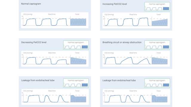
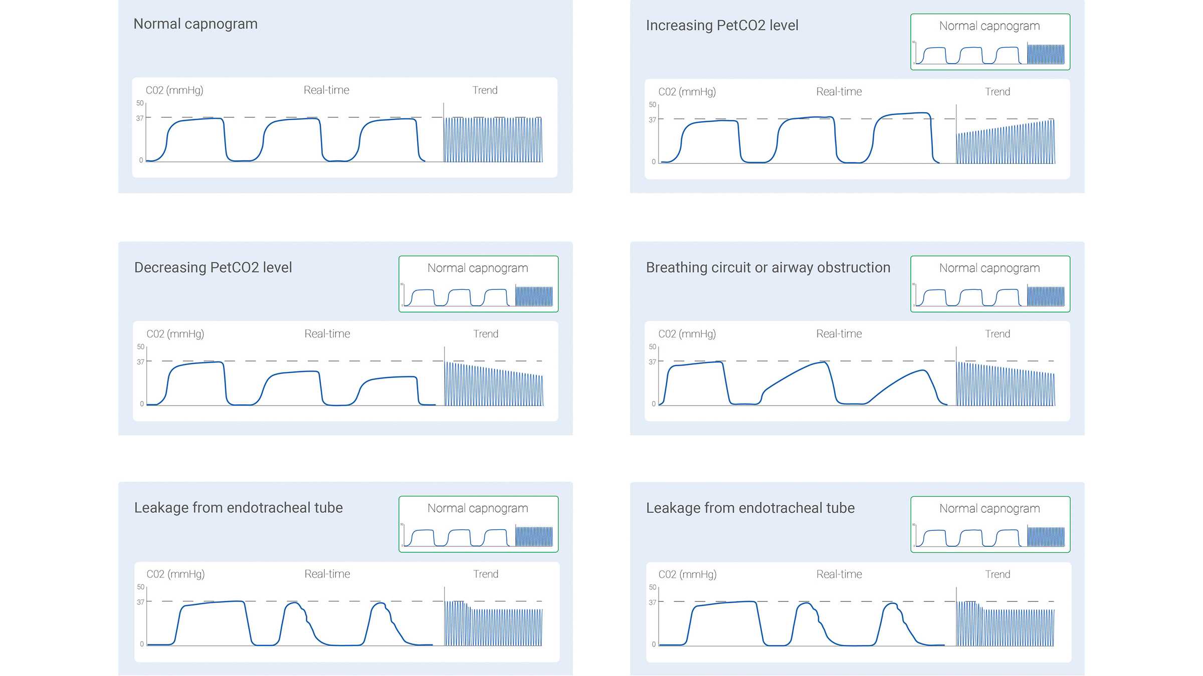
La señal de CO2 medida se puede registrar como una función de tiempo (capnografía temporal) o como volumen espirado (capnografía volumétrica). La cantidad de información que estos dos tipos de capnografía pueden proporcionar varía notablemente. En algunos estudios se han descrito algunas formas de los capnogramas temporales consideradas como típicas de situaciones clínicas específicas. En la figura 1 de abajo se muestran algunas de las más habituales.

Con todo, la capnografía temporal también tiene sus limitaciones: no puede dar una estimación precisa del estado de ventilación-perfusión de los pulmones, y tampoco se puede usar para calcular el componente de espacio muerto fisiológico. La capnografía volumétrica, pese a no ser tan sencilla y cómoda como la capnografía temporal, tiene la ventaja de que ofrece una cantidad de información considerablemente mayor.

Diagramas de capnogramas con estados habituales
Figura 1
Diagramas de capnogramas con estados habituales
Figura 1

El capnograma volumétrico: forma y fases

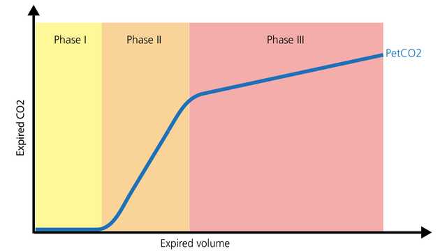
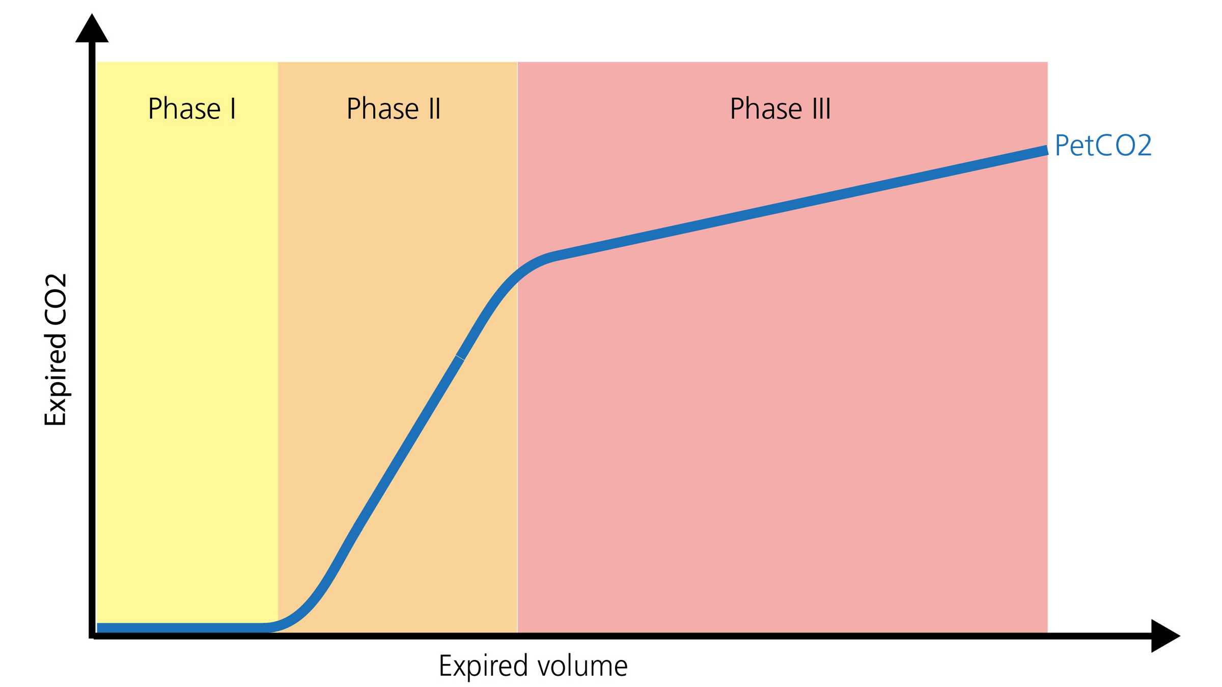
La forma normal de un capnograma volumétrico consta de tres fases. Es importante recordar que el capnograma representa una espiración.

  • La fase I representa el gas sin CO2 de las vías aéreas (espacio muerto anatómico instrumental).
  • La fase II es una fase de transición en la que el gas procedente de los conductos de aire se mezcla con gas alveolar.
  • La fase III es una fase meseta consistente en gas procedente de los alveolos y de las zonas pulmonares de vaciado lento (Gravenstein, J., Jaffe, M., & Paulus, D. (2004). Capnography: Clinical Aspects. New York: Cambridge University Press.2). En la figura 2 de abajo se muestra una representación visual.
Diagrama de las tres fases
Figura 2
Diagrama de las tres fases
Figura 2

Capnografía en los traslados

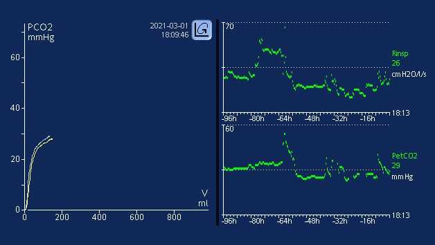
La capnografía, tanto si es temporal como volumétrica, puede arrojar información muy valiosa para optimizar la monitorización y guiar los cuidados de pacientes que deben trasladarse a otra zona del hospital o a otro hospital. Se puede utilizar de forma segura con tubos endotraqueales, tubos de traqueotomía y numerosas vías aéreas supraglóticas, siempre y cuando haya un sellado eficaz. La colocación y permeabilidad de la vía aérea, la monitorización de la ventilación y el estado de la perfusión son todos ámbitos en los que el valor de PetCO2 proporciona información significativa. Otro parámetro muy útil es el volumen de dióxido de carbono eliminado por minuto (V'CO2), que permite a los profesionales sanitarios valorar los esfuerzos efectivos de reanimación de volumen y perfusión (I-Gnaidy E., Abo El-Nasr, L., Ameen, S., & Abd El-Ghafar, M. (2019). Correlation between Cardon Dioxide Production and Mean Arterial Blood Pressure in Fluid Response in Mechanically Ventilated Patients. Medical Journal of Cairo University, 87(4), 2679-2684.3).

Capnografía en la UCI

En las unidades de cuidados intensivos, la capnografía de forma de onda permite seguir monitorizando la colocación y permeabilidad de la vía aérea, con los correspondientes dispositivos complementarios de vía aérea. La relación existente entre el espacio muerto anatómico y el volumen tidal (VD/Vt) es una medición de capnografía importante.  Una relación VD/Vt en aumento puede representar un posible incremento de la mortalidad dependiendo del nivel de aumento (Kallet RH, Alonso JA, Pittet JF, Matthay MA. Prognostic value of the pulmonary dead-space fraction during the first 6 days of acute respiratory distress syndrome. Respir Care. 2004;49(9):1008-1014. 4, Nuckton TJ, Alonso JA, Kallet RH, et al. Pulmonary dead-space fraction as a risk factor for death in the acute respiratory distress syndrome. N Engl J Med. 2002;346(17):1281-1286. doi:10.1056/NEJMoa0128355). Los profesionales sanitarios pueden utilizar la forma de onda de PetCO2 y V’CO2 para optimizar el reclutamiento pulmonar, validar los ajustes de PEEP óptimos y detectar problemas con la perfusión (sistémica y pulmonar) (Kallet RH, Alonso JA, Pittet JF, Matthay MA. Prognostic value of the pulmonary dead-space fraction during the first 6 days of acute respiratory distress syndrome. Respir Care. 2004;49(9):1008-1014. 4, Nuckton TJ, Alonso JA, Kallet RH, et al. Pulmonary dead-space fraction as a risk factor for death in the acute respiratory distress syndrome. N Engl J Med. 2002;346(17):1281-1286. doi:10.1056/NEJMoa0128355, Blankman P, Shono A, Hermans BJ, Wesselius T, Hasan D, Gommers D. Detection of optimal PEEP for equal distribution of tidal volume by volumetric capnography and electrical impedance tomography during decreasing levels of PEEP in post cardiac-surgery patients. Br J Anaesth. 2016;116(6):862-869. doi:10.1093/bja/aew1166, Nguyen LS, Squara P. Non-Invasive Monitoring of Cardiac Output in Critical Care Medicine. Front Med (Lausanne). 2017;4:200. Published 2017 Nov 20. doi:10.3389/fmed.2017.002007). El V’CO2 es un valor que también se puede usar durante la retirada de la ventilación mecánica, que permite a los profesionales sanitarios detectar posibles problemas o fatiga del paciente (mayor fracción del espacio muerto, un esfuerzo inadecuado y fatiga del músculo respiratorio). El gasto energético derivado del V’CO2 es un método preciso que los profesionales sanitarios pueden emplear para calcular los requisitos nutricionales de los pacientes con ventilación mecánica (Stapel SN, de Grooth HJ, Alimohamad H, et al. Ventilator-derived carbon dioxide production to assess energy expenditure in critically ill patients: proof of concept. Crit Care. 2015;19:370. Published 2015 Oct 22. doi:10.1186/s13054-015-1087-28).

Todos los respiradores de Hamilton Medical incorporan capnografía volumétrica (Todos los modelos salvo el HAMILTON-MR1A), ya sea de serie o como característica opcional. La medición del CO2 se lleva a cabo mediante un sensor de CO2 convencional, el CAPNOSTAT® 5, en la abertura de la vía aérea del paciente. Aparte, ofrecen una visión general de todos los valores de CO2 pertinentes en la ventana Monitorización de CO2.

Libro electrónico de capnografía volumétrica

Libro electrónico gratuito

¡Siga aprendiendo! Todo sobre la capnografía volumétrica

Aprenda a interpretar capnogramas volumétricos y obtenga una visión general de las ventajas y las aplicaciones clínicas de la capnografía volumétrica. Incluye una prueba de autoevaluación.

Notas al pie

  • A. Todos los modelos excepto HAMILTON-MR1

Referencias

  1. 1. Kremeier P, Böhm SH, Tusman G. Clinical use of volumetric capnography in mechanically ventilated patients. J Clin Monit Comput. 2020;34(1):7-16. doi:10.1007/s10877-019-00325-9
  2. 2. Gravenstein, J., Jaffe, M., & Paulus, D. (2004). Capnography: Clinical Aspects. New York: Cambridge University Press.
  3. 3. I-Gnaidy E., Abo El-Nasr, L., Ameen, S., & Abd El-Ghafar, M. (2019). Correlation between Cardon Dioxide Production and Mean Arterial Blood Pressure in Fluid Response in Mechanically Ventilated Patients. Medical Journal of Cairo University, 87(4), 2679-2684.
  4. 4. Kallet RH, Alonso JA, Pittet JF, Matthay MA. Prognostic value of the pulmonary dead-space fraction during the first 6 days of acute respiratory distress syndrome. Respir Care. 2004;49(9):1008-1014.
  5. 5. Nuckton TJ, Alonso JA, Kallet RH, et al. Pulmonary dead-space fraction as a risk factor for death in the acute respiratory distress syndrome. N Engl J Med. 2002;346(17):1281-1286. doi:10.1056/NEJMoa012835
  6. 6. Blankman P, Shono A, Hermans BJ, Wesselius T, Hasan D, Gommers D. Detection of optimal PEEP for equal distribution of tidal volume by volumetric capnography and electrical impedance tomography during decreasing levels of PEEP in post cardiac-surgery patients. Br J Anaesth. 2016;116(6):862-869. doi:10.1093/bja/aew116
  7. 7. Nguyen LS, Squara P. Non-Invasive Monitoring of Cardiac Output in Critical Care Medicine. Front Med (Lausanne). 2017;4:200. Published 2017 Nov 20. doi:10.3389/fmed.2017.00200
  8. 8. Stapel SN, de Grooth HJ, Alimohamad H, et al. Ventilator-derived carbon dioxide production to assess energy expenditure in critically ill patients: proof of concept. Crit Care. 2015;19:370. Published 2015 Oct 22. doi:10.1186/s13054-015-1087-2

Clinical use of volumetric capnography in mechanically ventilated patients.

Kremeier P, Böhm SH, Tusman G. Clinical use of volumetric capnography in mechanically ventilated patients. J Clin Monit Comput. 2020;34(1):7-16. doi:10.1007/s10877-019-00325-9

Capnography is a first line monitoring system in mechanically ventilated patients. Volumetric capnography supports noninvasive and breath-by-breath information at the bedside using mainstream CO2 and flow sensors placed at the airways opening. This volume-based capnography provides information of important body functions related to the kinetics of carbon dioxide. Volumetric capnography goes one step forward standard respiratory mechanics and provides a new dimension for monitoring of mechanical ventilation. The article discusses the role of volumetric capnography for the clinical monitoring of mechanical ventilation.

Capnography: Clinical Aspects

Gravenstein, J., Jaffe, M., & Paulus, D. (2004). Capnography: Clinical Aspects. New York: Cambridge University Press.

Correlation between Carbon Dioxide Production and Mean Arterial Blood Pressure in Fluid Response in Mechanically Ventilated Patients

I-Gnaidy E., Abo El-Nasr, L., Ameen, S., & Abd El-Ghafar, M. (2019). Correlation between Cardon Dioxide Production and Mean Arterial Blood Pressure in Fluid Response in Mechanically Ventilated Patients. Medical Journal of Cairo University, 87(4), 2679-2684.

Prognostic value of the pulmonary dead-space fraction during the first 6 days of acute respiratory distress syndrome.

Kallet RH, Alonso JA, Pittet JF, Matthay MA. Prognostic value of the pulmonary dead-space fraction during the first 6 days of acute respiratory distress syndrome. Respir Care. 2004;49(9):1008-1014.


BACKGROUND

The ratio of pulmonary dead space to tidal volume (VD/VT) in acute respiratory distress syndrome (ARDS) is reported to be between 0.35 and 0.55. However, VD/VT has seldom been measured with consideration to the evolving pathophysiology of ARDS.

METHODS

We made serial VD/VT measurements with 59 patients who required mechanical ventilation for > or = 6 days. We measured VD/VT within 24 h of the point at which the patient met the American-European Consensus Conference criteria for ARDS, and we repeated the VD/VT measurement on ARDS days 2, 3, and 6 with a bedside metabolic monitor during volume-regulated ventilation. We analyzed the changes in VD/VT over the 6-day period to determine whether VD/VT has a significant association with mortality.

RESULTS

VD/VT was significantly higher in nonsurvivors on day 1 (0.61 +/- 0.09 vs 0.54 +/- 0.08, p < 0.05), day 2 (0.63 +/- 0.09 vs 0.53 +/- 0.09, p < 0.001), day 3 (0.64 +/- 0.09 vs 0.53 +/- 0.09, p < 0.001), and day 6 (0.66 +/- 0.09 vs 0.51 +/- 0.08, p < 0.001).

CONCLUSION

In ARDS a sustained VD/VT elevation is characteristic of nonsurvivors, so dead-space measurements made beyond the first 24 hours may have prognostic value.

Pulmonary dead-space fraction as a risk factor for death in the acute respiratory distress syndrome.

Nuckton TJ, Alonso JA, Kallet RH, et al. Pulmonary dead-space fraction as a risk factor for death in the acute respiratory distress syndrome. N Engl J Med. 2002;346(17):1281-1286. doi:10.1056/NEJMoa012835


BACKGROUND

No single pulmonary-specific variable, including the severity of hypoxemia, has been found to predict the risk of death independently when measured early in the course of the acute respiratory distress syndrome. Because an increase in the pulmonary dead-space fraction has been described in observational studies of the syndrome, we systematically measured the dead-space fraction early in the course of the illness and evaluated its potential association with the risk of death.

METHODS

The dead-space fraction was prospectively measured in 179 intubated patients, a mean (+/-SD) of 10.9+/-7.4 hours after the acute respiratory distress syndrome had developed. Additional clinical and physiological variables were analyzed with the use of multiple logistic regression. The study outcome was mortality before hospital discharge.

RESULTS

The mean dead-space fraction was markedly elevated (0.58+/-0.09) early in the course of the acute respiratory distress syndrome and was higher among patients who died than among those who survived (0.63+/-0.10 vs. 0.54+/-0.09, P<0.001). The dead-space fraction was an independent risk factor for death: for every 0.05 increase, the odds of death increased by 45 percent (odds ratio, 1.45; 95 percent confidence interval, 1.15 to 1.83; P=0.002). The only other independent predictors of an increased risk of death were the Simplified Acute Physiology Score II, an indicator of the severity of illness (odds ratio, 1.06; 95 percent confidence interval, 1.03 to 1.08; P<0.001) and quasistatic respiratory compliance (odds ratio, 1.06; 95 percent confidence interval, 1.01 to 1.10; P=0.01).

CONCLUSIONS

Increased dead-space fraction is a feature of the early phase of the acute respiratory distress syndrome. Elevated values are associated with an increased risk of death.

Detection of optimal PEEP for equal distribution of tidal volume by volumetric capnography and electrical impedance tomography during decreasing levels of PEEP in post cardiac-surgery patients.

Blankman P, Shono A, Hermans BJ, Wesselius T, Hasan D, Gommers D. Detection of optimal PEEP for equal distribution of tidal volume by volumetric capnography and electrical impedance tomography during decreasing levels of PEEP in post cardiac-surgery patients. Br J Anaesth. 2016;116(6):862-869. doi:10.1093/bja/aew116


BACKGROUND

Homogeneous ventilation is important for prevention of ventilator-induced lung injury. Electrical impedance tomography (EIT) has been used to identify optimal PEEP by detection of homogenous ventilation in non-dependent and dependent lung regions. We aimed to compare the ability of volumetric capnography and EIT in detecting homogenous ventilation between these lung regions.

METHODS

Fifteen mechanically-ventilated patients after cardiac surgery were studied. Ventilator settings were adjusted to volume-controlled mode with a fixed tidal volume (Vt) of 6-8 ml kg(-1) predicted body weight. Different PEEP levels were applied (14 to 0 cm H2O, in steps of 2 cm H2O) and blood gases, Vcap and EIT were measured.

RESULTS

Tidal impedance variation of the non-dependent region was highest at 6 cm H2O PEEP, and decreased significantly at 14 cm H2O PEEP indicating decrease in the fraction of Vt in this region. At 12 cm H2O PEEP, homogenous ventilation was seen between both lung regions. Bohr and Enghoff dead space calculations decreased from a PEEP of 10 cm H2O. Alveolar dead space divided by alveolar Vt decreased at PEEP levels ≤6 cm H2O. The normalized slope of phase III significantly changed at PEEP levels ≤4 cm H2O. Airway dead space was higher at higher PEEP levels and decreased at the lower PEEP levels.

CONCLUSIONS

In postoperative cardiac patients, calculated dead space agreed well with EIT to detect the optimal PEEP for an equal distribution of inspired volume, amongst non-dependent and dependent lung regions. Airway dead space reduces at decreasing PEEP levels.

Non-Invasive Monitoring of Cardiac Output in Critical Care Medicine.

Nguyen LS, Squara P. Non-Invasive Monitoring of Cardiac Output in Critical Care Medicine. Front Med (Lausanne). 2017;4:200. Published 2017 Nov 20. doi:10.3389/fmed.2017.00200

Critically ill patients require close hemodynamic monitoring to titrate treatment on a regular basis. It allows administering fluid with parsimony and adjusting inotropes and vasoactive drugs when necessary. Although invasive monitoring is considered as the reference method, non-invasive monitoring presents the obvious advantage of being associated with fewer complications, at the expanse of accuracy, precision, and step-response change. A great many methods and devices are now used over the world, and this article focuses on several of them, providing with a brief review of related underlying physical principles and validation articles analysis. Reviewed methods include electrical bioimpedance and bioreactance, respiratory-derived cardiac output (CO) monitoring technique, pulse wave transit time, ultrasound CO monitoring, multimodal algorithmic estimation, and inductance thoracocardiography. Quality criteria with which devices were reviewed included: accuracy (closeness of agreement between a measurement value and a true value of the measured), precision (closeness of agreement between replicate measurements on the same or similar objects under specified conditions), and step response change (delay between physiological change and its indication). Our conclusion is that the offer of non-invasive monitoring has improved in the past few years, even though further developments are needed to provide clinicians with sufficiently accurate devices for routine use, as alternative to invasive monitoring devices.

Ventilator-derived carbon dioxide production to assess energy expenditure in critically ill patients: proof of concept.

Stapel SN, de Grooth HJ, Alimohamad H, et al. Ventilator-derived carbon dioxide production to assess energy expenditure in critically ill patients: proof of concept. Crit Care. 2015;19:370. Published 2015 Oct 22. doi:10.1186/s13054-015-1087-2


INTRODUCTION

Measurement of energy expenditure (EE) is recommended to guide nutrition in critically ill patients. Availability of a gold standard indirect calorimetry is limited, and continuous measurement is unfeasible. Equations used to predict EE are inaccurate. The purpose of this study was to provide proof of concept that EE can be accurately assessed on the basis of ventilator-derived carbon dioxide production (VCO2) and to determine whether this method is more accurate than frequently used predictive equations.

METHODS

In 84 mechanically ventilated critically ill patients, we performed 24-h indirect calorimetry to obtain a gold standard EE. Simultaneously, we collected 24-h ventilator-derived VCO2, extracted the respiratory quotient of the administered nutrition, and calculated EE with a rewritten Weir formula. Bias, precision, and accuracy and inaccuracy rates were determined and compared with four predictive equations: the Harris-Benedict, Faisy, and Penn State University equations and the European Society for Clinical Nutrition and Metabolism (ESPEN) guideline equation of 25 kcal/kg/day.

RESULTS

Mean 24-h indirect calorimetry EE was 1823 ± 408 kcal. EE from ventilator-derived VCO2 was accurate (bias +141 ± 153 kcal/24 h; 7.7 % of gold standard) and more precise than the predictive equations (limits of agreement -166 to +447 kcal/24 h). The 10 % and 15 % accuracy rates were 61 % and 76 %, respectively, which were significantly higher than those of the Harris-Benedict, Faisy, and ESPEN guideline equations. Large errors of more than 30 % inaccuracy did not occur with EE derived from ventilator-derived VCO2. This 30 % inaccuracy rate was significantly lower than that of the predictive equations.

CONCLUSIONS

In critically ill mechanically ventilated patients, assessment of EE based on ventilator-derived VCO2 is accurate and more precise than frequently used predictive equations. It allows for continuous monitoring and is the best alternative to indirect calorimetry.

Related articles. Get a deeper look