Atrás

Cómo medir la presión esofágica correctamente

Artículo

Autor: Jean-Michel Arnal, intensivista jefe del Hôpital Sainte Musse, Toulon, Francia

Fecha: 19.10.2018

Last change: 09.09.2020

Formatting

Un estudio fisiológico reciente pone de manifiesto que la presión esofágica permite estimar la presión pleural en la región central del tórax en todos los niveles de PEEP. Por tanto, una medición absoluta de la presión esofágica resulta útil a la hora de establecer la PEEP y de monitorizar la presión transpulmonar.

Cómo medir la presión esofágica correctamente

Colocación inicial e inflado

Entonces, ¿cómo se mide la presión esofágica correctamente?

El globo esofágico debe colocarse e inflarse de forma adecuada, y su colocación debe verificarse.

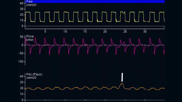
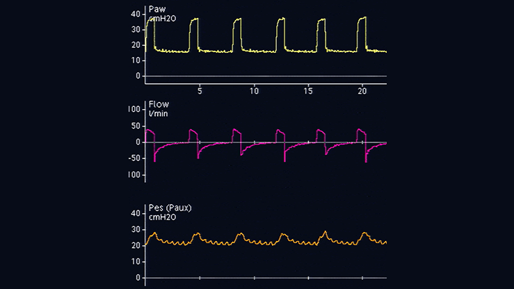
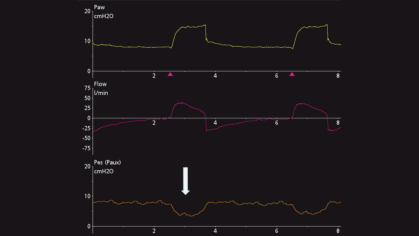
La colocación óptima del globo esofágico es el tercio inferior del esófago, a una distancia de 35–45 cm de las fosas nasales. Con un paciente en posición parcialmente decúbito, el globo vacío se introduce en el estómago, que está a unos 50–60 cm de las fosas nasales. El globo se infla a un volumen estándar (1 ml con un catéter CooperSurgical y 4 ml con un catéter Nutrivent). La presión gástrica muestra un desvío positivo durante la inspiración en pacientes con respiración tanto espontánea como pasiva. La colocación gástrica se determina ejerciendo una suave compresión epigástrica manual, que produce un aumento inmediato de la presión gástrica (consulte la figura 1).

Captura de pantalla de formas de onda que revelan un aumento de la Pes
Figura 1
Captura de pantalla de formas de onda que revelan un aumento de la Pes
Figura 1

Retirada con el globo inflado

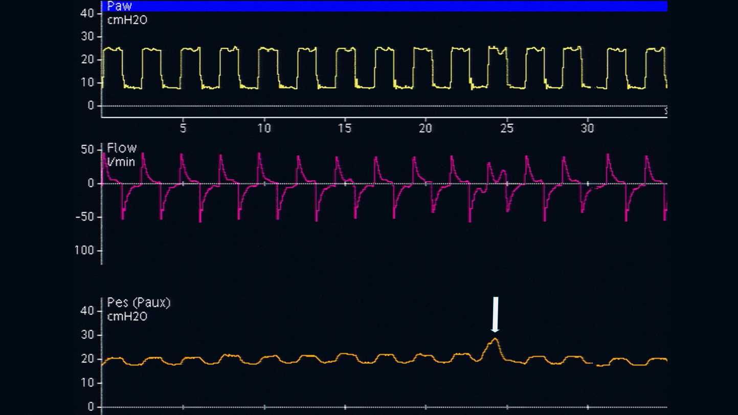
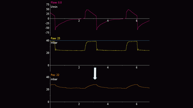
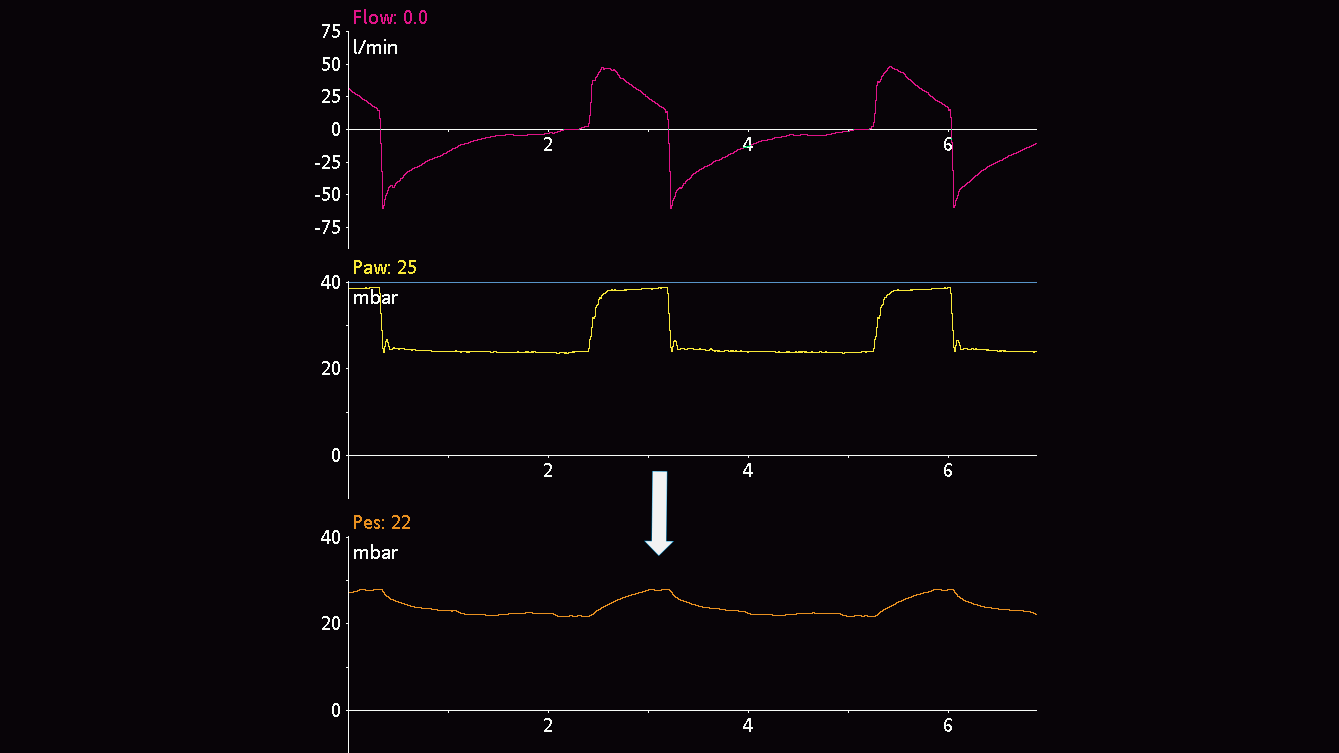
A continuación, el catéter esofágico se va retirando suavemente mientras el globo se infla, lo que ayudará a colocar el globo en el tercio inferior del esófago. Durante el cambio de presión gástrica (consulte la figura 2) a esofágica (consulte la figura 3), la línea de referencia de la forma de onda de presión cambia y aparecen oscilaciones cardiacas.

Forma de onda que muestra la línea de referencia de la presión gástrica
Figura 2: presión gástrica
Forma de onda que muestra la línea de referencia de la presión gástrica
Figura 2: presión gástrica
Forma de onda que muestra la línea de referencia de la presión esofágica
Figura 3: presión esofágica
Forma de onda que muestra la línea de referencia de la presión esofágica
Figura 3: presión esofágica

Desvíos de la presión esofágica

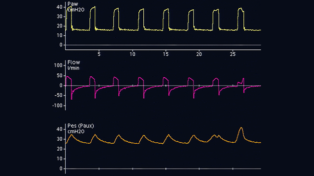
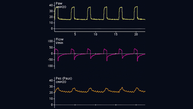
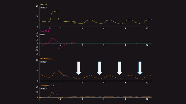
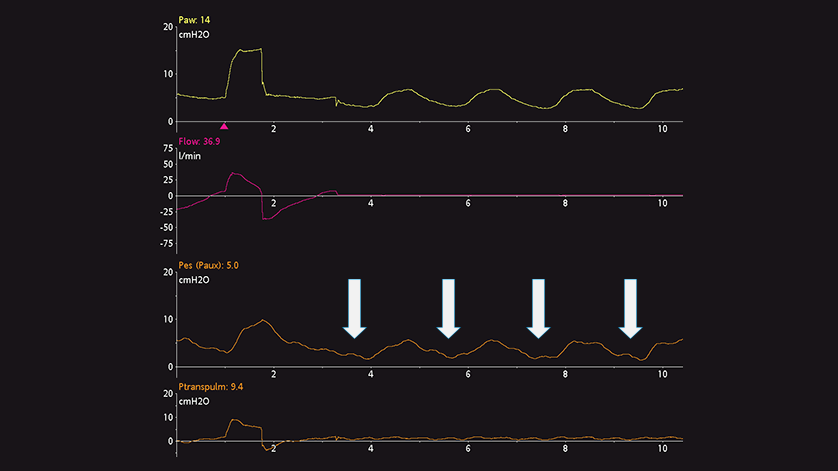
Los desvíos en la presión esofágica son positivos durante la inspiración en los pacientes pasivos (consulte la figura 4), pero negativos en el caso de los pacientes con respiración espontánea (consulte la figura 5). Si se producen oscilaciones cardiacas que distorsionan la señal de presión esofágica, el catéter se puede retirar unos 2–5 cm más.
 

Forma de onda que muestra desvíos de la Pes en un paciente con respiración pasiva
Figura 4: paciente pasivo
Forma de onda que muestra desvíos de la Pes en un paciente con respiración pasiva
Figura 4: paciente pasivo
Forma de onda que muestra desvíos de la Pes en un paciente con respiración espontánea
Figura 5: paciente con respiración espontánea
Forma de onda que muestra desvíos de la Pes en un paciente con respiración espontánea
Figura 5: paciente con respiración espontánea

Inflado del globo

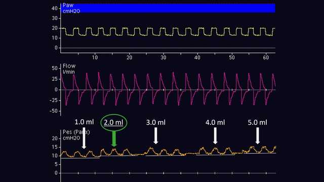
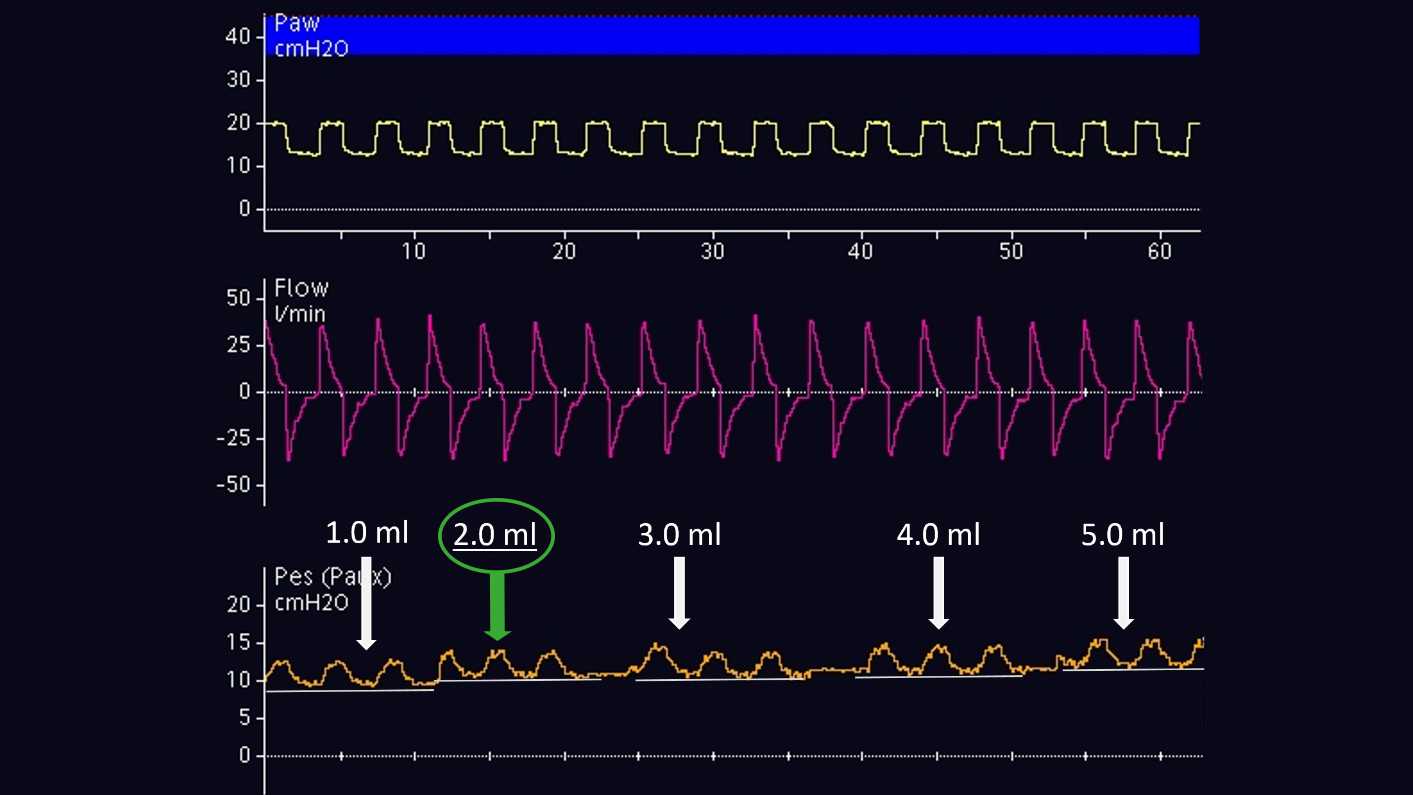
El volumen de aire para lograr el inflado adecuado del globo se debe valorar individualmente. Esto solo es posible en los pacientes pasivos. Según el método propuesto por Mojoli et al (2016), el globo se infla entre 0,5 y 3 ml a intervalos de 0,5 ml con un catéter CooperSurgical, y entre 1 y 8 ml a intervalos de 1 ml con un catéter Nutrivent (consulte la figura 6). Durante el inflado progresivo del globo, la línea de referencia de la presión esofágica aumenta y la magnitud del desvío de la presión esofágica cambia. El volumen de inflado adecuado es el que está asociado al desvío más pronunciado de la presión esofágica. Si dos volúmenes distintos muestran la misma magnitud de desvío de la presión esofágica, se seleccionará el volumen más bajo.

Forma de onda durante el inflado de un globo
Figura 6: inflado de globo realizado con un catéter Nutrivent
Forma de onda durante el inflado de un globo
Figura 6: inflado de globo realizado con un catéter Nutrivent

Verificación

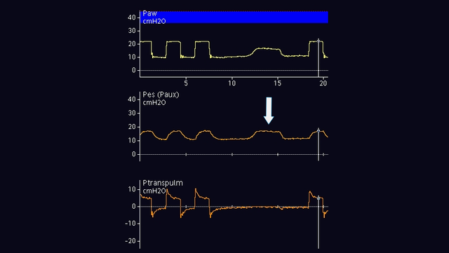
Una vez que el globo está correctamente colocado en el esófago y se ha inflado, se debe realizar una verificación mediante una prueba de oclusión. El principio consiste en cerrar las vías aéreas al final de la espiración para cambiar la presión en estas y, luego, confirmar que la presión esofágica cambia en la misma medida.

En los pacientes con respiración pasiva, se puede efectuar una oclusión al final de la espiración. Cuando la válvula espiratoria se cierre, ejerza una presión manual externa en la caja torácica, a ambos lados del pecho, para ver un desvío positivo de las presiones en la vía aérea y esofágica. La magnitud del aumento de las presiones en la vía aérea y esofágica debe ser la misma. Dicho de otro modo, la presión transpulmonar debe permanecer inalterada (consulte la figura 7).

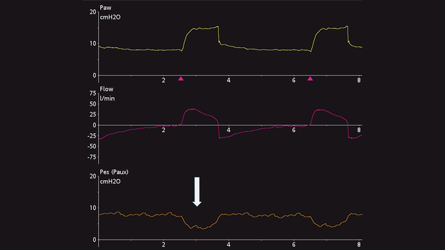
En los pacientes con respiración activa, la prueba de oclusión dinámica también emplea una oclusión al final de la espiración. No es necesario ejercer ninguna presión manual sobre el tórax, ya que el paciente realizará el esfuerzo inspiratorio espontáneo durante la oclusión. El resultado es un desvío negativo de las presiones en la vía aérea y esofágica. La magnitud de la reducción de las presiones en la vía aérea y esofágica debe ser la misma, esto es, la presión transpulmonar debe permanecer inalterada (consulte la figura 8).

Si desea monitorizar la presión esofágica constantemente, es importante reevaluar la correcta colocación y el volumen de inflado.

Puede consultar las citas completas a continuación: (Yoshida T, Amato MBP, Grieco DL, et al. Esophageal Manometry and Regional Transpulmonary Pressure in Lung Injury. Am J Respir Crit Care Med. 2018;197(8):1018-1026. doi:10.1164/rccm.201709-1806OC1, Mojoli F, Iotti GA, Torriglia F, et al. In vivo calibration of esophageal pressure in the mechanically ventilated patient makes measurements reliable. Crit Care. 2016;20:98. Published 2016 Apr 11. doi:10.1186/s13054-016-1278-52, Baydur A, Behrakis PK, Zin WA, Jaeger M, Milic-Emili J. A simple method for assessing the validity of the esophageal balloon technique. Am Rev Respir Dis. 1982;126(5):788-791. doi:10.1164/arrd.1982.126.5.7883, Akoumianaki E, Maggiore SM, Valenza F, et al. The application of esophageal pressure measurement in patients with respiratory failure. Am J Respir Crit Care Med. 2014;189(5):520-531. doi:10.1164/rccm.201312-2193CI4, Mauri T, Yoshida T, Bellani G, et al. Esophageal and transpulmonary pressure in the clinical setting: meaning, usefulness and perspectives. Intensive Care Med. 2016;42(9):1360-1373. doi:10.1007/s00134-016-4400-x5)

Forma de onda que muestra un aumento de Pva y Pes; sin cambios en Ptranspulm
Figura 7: prueba de oclusión en paciente con respiración pasiva
Forma de onda que muestra un aumento de Pva y Pes; sin cambios en Ptranspulm
Figura 7: prueba de oclusión en paciente con respiración pasiva
Forma de onda que muestra una reducción de Pva y Pes; sin cambios en Ptranspulm
Figura 8: prueba de oclusión en paciente con respiración activa
Forma de onda que muestra una reducción de Pva y Pes; sin cambios en Ptranspulm
Figura 8: prueba de oclusión en paciente con respiración activa
Libro electrónico con 13 consejos de expertos

Libro electrónico gratuito

13 consejos de expertos. Medición de la presión esofágica

Recomendaciones probadas clínicamente sobre qué hacer y qué evitar a la hora de usar la medición de la presión esofágica en pacientes con SDRA.

Esophageal Manometry and Regional Transpulmonary Pressure in Lung Injury.

Yoshida T, Amato MBP, Grieco DL, et al. Esophageal Manometry and Regional Transpulmonary Pressure in Lung Injury. Am J Respir Crit Care Med. 2018;197(8):1018-1026. doi:10.1164/rccm.201709-1806OC


RATIONALE

Esophageal manometry is the clinically available method to estimate pleural pressure, thus enabling calculation of transpulmonary pressure (Pl). However, many concerns make it uncertain in which lung region esophageal manometry reflects local Pl.

OBJECTIVES

To determine the accuracy of esophageal pressure (Pes) and in which regions esophageal manometry reflects pleural pressure (Ppl) and Pl; to assess whether lung stress in nondependent regions can be estimated at end-inspiration from Pl.

METHODS

In lung-injured pigs (n = 6) and human cadavers (n = 3), Pes was measured across a range of positive end-expiratory pressure, together with directly measured Ppl in nondependent and dependent pleural regions. All measurements were obtained with minimal nonstressed volumes in the pleural sensors and esophageal balloons. Expiratory and inspiratory Pl was calculated by subtracting local Ppl or Pes from airway pressure; inspiratory Pl was also estimated by subtracting Ppl (calculated from chest wall and respiratory system elastance) from the airway plateau pressure.

MEASUREMENTS AND MAIN RESULTS

In pigs and human cadavers, expiratory and inspiratory Pl using Pes closely reflected values in dependent to middle lung (adjacent to the esophagus). Inspiratory Pl estimated from elastance ratio reflected the directly measured nondependent values.

CONCLUSIONS

These data support the use of esophageal manometry in acute respiratory distress syndrome. Assuming correct calibration, expiratory Pl derived from Pes reflects Pl in dependent to middle lung, where atelectasis usually predominates; inspiratory Pl estimated from elastance ratio may indicate the highest level of lung stress in nondependent "baby" lung, where it is vulnerable to ventilator-induced lung injury.

In vivo calibration of esophageal pressure in the mechanically ventilated patient makes measurements reliable.

Mojoli F, Iotti GA, Torriglia F, et al. In vivo calibration of esophageal pressure in the mechanically ventilated patient makes measurements reliable. Crit Care. 2016;20:98. Published 2016 Apr 11. doi:10.1186/s13054-016-1278-5

In screening programmes it is important to assess a preliminary effectiveness of the screening method as soon as possible in order to forecast survival figures. In March 1981 a controlled single-view mammographic screening trial for breast cancer was started in the south of Stockholm. The population invited for screening mammography consisted of 40,000 women aged 40-64 years, and 20,000 women served as a well-defined control group. The main aim of the trial was to determine whether repeated mammographic screening could reduce the mortality in the study population (SP) compared to the control population (CP). The cumulative number of advanced mammary carcinomas in the screening and the control populations from the first five years of screening have shown a tendency towards more favourable stages in the screened population aged 40-64 years. A breakdown by age suggests an effect in age group 50-59 years, but not yet in age groups 40-49 and 60-64 years. When comparing the rates of stage II+ cancer, an increased number is found in the study group. As the total rate of breast cancer is higher in SP than in CP, there ought to be a concealed group of stage II+ cancers in the CP which makes the comparison biased. A new approach has been designed, where an estimation of the 'hidden' number of stage II+ cancers in CP is added to the clinically detected cases, and in this respect a comparison has shown a decrease in the cumulative number of advanced cancers in the SP in relation to the CP (p less than 0.05).(ABSTRACT TRUNCATED AT 250 WORDS)

A simple method for assessing the validity of the esophageal balloon technique.

Baydur A, Behrakis PK, Zin WA, Jaeger M, Milic-Emili J. A simple method for assessing the validity of the esophageal balloon technique. Am Rev Respir Dis. 1982;126(5):788-791. doi:10.1164/arrd.1982.126.5.788

The validity of the conventional esophageal balloon technique as a measure of pleural pressure was tested in 10 subjects in sitting, supine, and lateral positions by occluding the airways at end-expiration and measuring the ratio of changes in esophageal (delta Pes) and mouth pressure (delta Pm) during the ensuing spontaneous occluded inspiratory efforts. Similar measurements were also made during static Mueller maneuvers. In both tests, delta Pes/delta Pm values were close to unity in sitting and lateral positions, whereas in the supine position, substantial deviations from unity were found in some instances. However, by repositioning the balloon to different levels in the esophagus, even in these instances a locus could be found where the delta Pes/delta Pm ratio was close to unity. No appreciable phase difference between delta Pes and delta Pm was found. We conclude that by positioning the balloon according to the "occlusion test" procedure, valid measurements of pleural pressure can be obtained in all the tested body positions.

The application of esophageal pressure measurement in patients with respiratory failure.

Akoumianaki E, Maggiore SM, Valenza F, et al. The application of esophageal pressure measurement in patients with respiratory failure. Am J Respir Crit Care Med. 2014;189(5):520-531. doi:10.1164/rccm.201312-2193CI

This report summarizes current physiological and technical knowledge on esophageal pressure (Pes) measurements in patients receiving mechanical ventilation. The respiratory changes in Pes are representative of changes in pleural pressure. The difference between airway pressure (Paw) and Pes is a valid estimate of transpulmonary pressure. Pes helps determine what fraction of Paw is applied to overcome lung and chest wall elastance. Pes is usually measured via a catheter with an air-filled thin-walled latex balloon inserted nasally or orally. To validate Pes measurement, a dynamic occlusion test measures the ratio of change in Pes to change in Paw during inspiratory efforts against a closed airway. A ratio close to unity indicates that the system provides a valid measurement. Provided transpulmonary pressure is the lung-distending pressure, and that chest wall elastance may vary among individuals, a physiologically based ventilator strategy should take the transpulmonary pressure into account. For monitoring purposes, clinicians rely mostly on Paw and flow waveforms. However, these measurements may mask profound patient-ventilator asynchrony and do not allow respiratory muscle effort assessment. Pes also permits the measurement of transmural vascular pressures during both passive and active breathing. Pes measurements have enhanced our understanding of the pathophysiology of acute lung injury, patient-ventilator interaction, and weaning failure. The use of Pes for positive end-expiratory pressure titration may help improve oxygenation and compliance. Pes measurements make it feasible to individualize the level of muscle effort during mechanical ventilation and weaning. The time is now right to apply the knowledge obtained with Pes to improve the management of critically ill and ventilator-dependent patients.

Esophageal and transpulmonary pressure in the clinical setting: meaning, usefulness and perspectives.

Mauri T, Yoshida T, Bellani G, et al. Esophageal and transpulmonary pressure in the clinical setting: meaning, usefulness and perspectives. Intensive Care Med. 2016;42(9):1360-1373. doi:10.1007/s00134-016-4400-x


PURPOSE

Esophageal pressure (Pes) is a minimally invasive advanced respiratory monitoring method with the potential to guide management of ventilation support and enhance specific diagnoses in acute respiratory failure patients. To date, the use of Pes in the clinical setting is limited, and it is often seen as a research tool only.

METHODS

This is a review of the relevant technical, physiological and clinical details that support the clinical utility of Pes.

RESULTS

After appropriately positioning of the esophageal balloon, Pes monitoring allows titration of controlled and assisted mechanical ventilation to achieve personalized protective settings and the desired level of patient effort from the acute phase through to weaning. Moreover, Pes monitoring permits accurate measurement of transmural vascular pressure and intrinsic positive end-expiratory pressure and facilitates detection of patient-ventilator asynchrony, thereby supporting specific diagnoses and interventions. Finally, some Pes-derived measures may also be obtained by monitoring electrical activity of the diaphragm.

CONCLUSIONS

Pes monitoring provides unique bedside measures for a better understanding of the pathophysiology of acute respiratory failure patients. Including Pes monitoring in the intensivist's clinical armamentarium may enhance treatment to improve clinical outcomes.