Atrás

Cómo medir la presión de trabajo en pacientes con ventilación asistida

Artículo

Autor: Jean-Michel Arnal, Hôpital Sainte Musse, Toulon, Francia

Fecha: 18.09.2025

La presión de trabajo estática debe monitorizarse sistemáticamente a pie de cama, pero ¿cómo se mide?

¿Qué es la presión de trabajo estática?

En pacientes que reciben ventilación mecánica en los modos de asistencia, la presión de trabajo estática (∆Pstat) se asocia de forma independiente con la supervivencia en la UCI (Bellani G, Grassi A, Sosio S, et al. Driving Pressure Is Associated with Outcome during Assisted Ventilation in Acute Respiratory Distress Syndrome. Anesthesiology. 2019;131(3):594-604. doi:10.1097/ALN.00000000000028461, Grassi A, Bianchi I, Teggia Droghi M, et al. Increased Driving Pressure During Assisted Ventilation for Hypoxemic Respiratory Failure Is Associated with Lower ICU Survival: The ICEBERG Study. Am J Respir Crit Care Med. Published online June 20, 2025. doi:10.1164/rccm.202411-2146OC2). Por lo tanto, los médicos deben monitorizar sistemáticamente la ∆Pstat a pie de cama para asegurarse de que la respiración espontánea es segura y, de este modo, evitar que el paciente sufra lesiones pulmonares autoinfligidas. La ∆Pstat representa el cambio en la presión elástica aplicado al sistema respiratorio durante la respiración y se calcula como la presión meseta (Pmeseta) menos la PEEP.

Cómo se obtiene la presión meseta

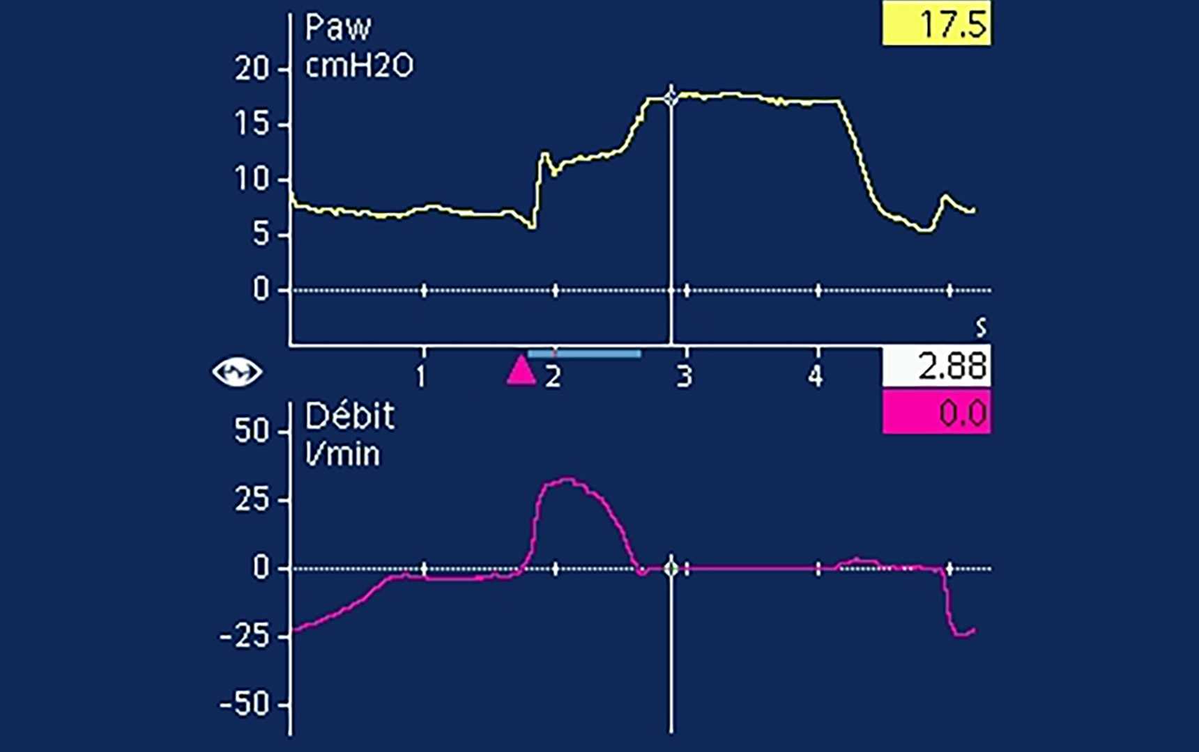
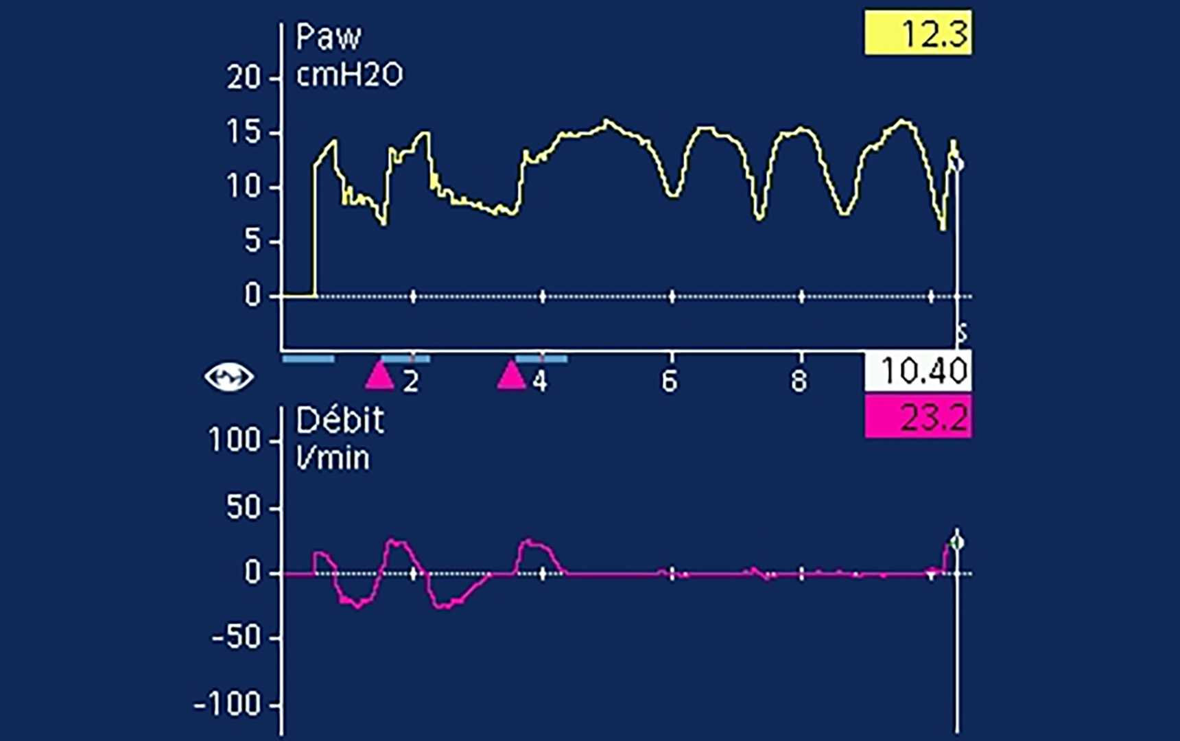
En las respiraciones asistidas, se utiliza una oclusión al final de la inspiración para medir la Pmeseta (Foti G, Cereda M, Banfi G, Pelosi P, Fumagalli R, Pesenti A. End-inspiratory airway occlusion: a method to assess the pressure developed by inspiratory muscles in patients with acute lung injury undergoing pressure support. Am J Respir Crit Care Med. 1997;156(4 Pt 1):1210-1216. Doi:10.1164/ajrccm.156.4.96-020313), para lo que debe identificarse visualmente una meseta de presión en las vías aéreas. En el caso de las respiraciones asistidas, la pausa al final de la inspiración se traduce en un aumento de la presión en las vías aéreas, que va seguida de una meseta (Bellani G, Grassi A, Sosio S, Foti G. Plateau and driving pressure in the presence of spontaneous breathing. Intensive Care Med. 2019;45(1):97-98. doi:10.1007/s00134-018-5311-94). El aumento de la presión en las vías aéreas hasta la Pmeseta se corresponde con el esfuerzo inspiratorio del paciente durante la respiración y se denomina índice de Pmus (PMI) (Foti G, Cereda M, Banfi G, Pelosi P, Fumagalli R, Pesenti A. End-inspiratory airway occlusion: a method to assess the pressure developed by inspiratory muscles in patients with acute lung injury undergoing pressure support. Am J Respir Crit Care Med. 1997;156(4 Pt 1):1210-1216. doi:10.1164/ajrccm.156.4.96-020313). Una presión meseta legible debe ir precedida de una rampa pronunciada al comienzo de la pausa, rápidamente seguida de una parte plana (figura 1) (Bianchi I, Grassi A, Pham T, et al. Reliability of plateau pressure during patient-triggered assisted ventilation. Analysis of a multicentre database. J Crit Care. 2022;68:96-103. doi:10.1016/j.jcrc.2021.12.0025). Los estudios observacionales realizados han notificado que la Pmeseta es legible en más del 90 % de las pausas (Bellani G, Grassi A, Sosio S, et al. Driving Pressure Is Associated with Outcome during Assisted Ventilation in Acute Respiratory Distress Syndrome. Anesthesiology. 2019;131(3):594-604. doi:10.1097/ALN.00000000000028461, Bianchi I, Grassi A, Pham T, et al. Reliability of plateau pressure during patient-triggered assisted ventilation. Analysis of a multicentre database. J Crit Care. 2022;68:96-103. doi:10.1016/j.jcrc.2021.12.0025). Las pausas no son legibles en aquellos casos en que no se puede determinar visualmente una meseta o cuando la actividad de los músculos inspiratorios y espiratorios se produce durante la pausa (figura 2) (Bellani G, Grassi A, Sosio S, et al. Driving Pressure Is Associated with Outcome during Assisted Ventilation in Acute Respiratory Distress Syndrome. Anesthesiology. 2019;131(3):594-604. doi:10.1097/ALN.00000000000028461).

Captura de pantalla donde se muestran las formas de onda de presión y de flujo con una meseta visible en la forma de onda de flujo
Figura 1: Una meseta visible se identifica con una Pmeseta de 17 cmH2O. En este paciente, la Ppico es de 12 cmH2O y la PEEP es de 7 cmH2O. Por lo tanto, el valor de ∆Pmeseta es 10 cmH2O y el del PMI, 5 cmH2O.
Captura de pantalla donde se muestra una presión meseta no legible
Figura 2a: Presión meseta no legible debido a la actividad de los músculos inspiratorios
Captura de pantalla donde se muestra una presión meseta no legible
Figura 2a: Presión meseta no legible debido a la actividad de los músculos inspiratorios
Captura de pantalla donde se muestra una presión meseta no legible debido a la actividad de los músculos espiratorios
Figura 2b: Presión meseta no legible debido a la actividad de los músculos espiratorios
Captura de pantalla donde se muestra una presión meseta no legible debido a la actividad de los músculos espiratorios
Figura 2b: Presión meseta no legible debido a la actividad de los músculos espiratorios
Captura de pantalla donde se muestra una presión meseta no legible debido a la actividad de los músculos espiratorios
Figura 2c: Presión meseta no legible debido a una combinación de ambas
Captura de pantalla donde se muestra una presión meseta no legible debido a la actividad de los músculos espiratorios
Figura 2c: Presión meseta no legible debido a una combinación de ambas

Cuidado con la sobrestimación

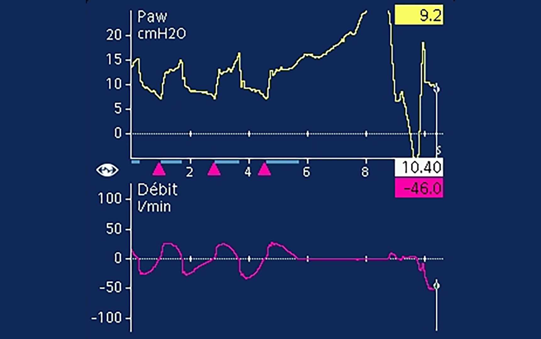
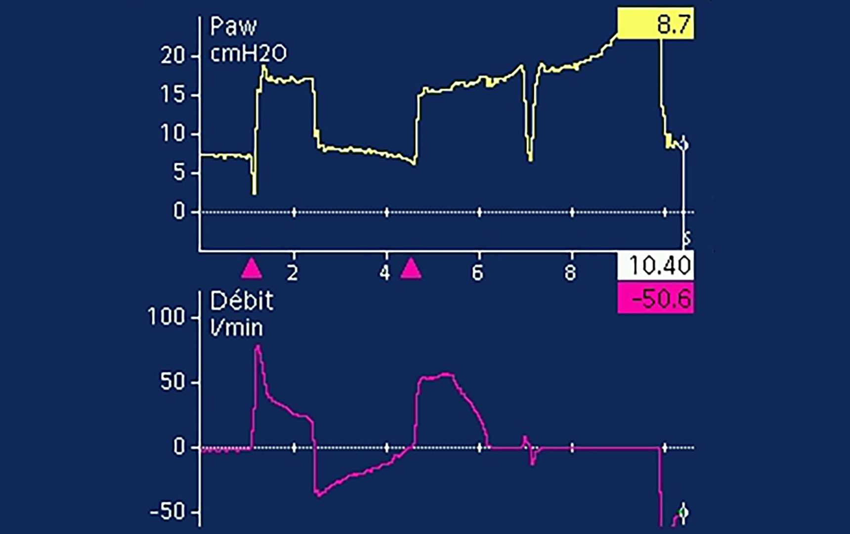
Incluso cuando la meseta es claramente visible, es frecuente que haya actividad de los músculos espiratorios presente (Soundoulounaki S, Akoumianaki E, Kondili E, et al. Airway pressure morphology and respiratory muscle activity during end-inspiratory occlusions in pressure support ventilation. Crit Care. 2020;24(1):467. Published 2020 Jul 28. doi:10.1186/s13054-020-03169-x6), lo que significa que la ∆Pstat puede sobrestimarse en las respiraciones asistidas. Por lo tanto, una ∆Pstat baja puede considerarse como segura.

Notas al pie

Referencias

  1. 1. Bellani G, Grassi A, Sosio S, et al. Driving Pressure Is Associated with Outcome during Assisted Ventilation in Acute Respiratory Distress Syndrome. Anesthesiology. 2019;131(3):594-604. doi:10.1097/ALN.0000000000002846
  2. 2. Grassi A, Bianchi I, Teggia Droghi M, et al. Increased Driving Pressure During Assisted Ventilation for Hypoxemic Respiratory Failure Is Associated with Lower ICU Survival: The ICEBERG Study. Am J Respir Crit Care Med. Published online June 20, 2025. doi:10.1164/rccm.202411-2146OC
  3. 3. Foti G, Cereda M, Banfi G, Pelosi P, Fumagalli R, Pesenti A. End-inspiratory airway occlusion: a method to assess the pressure developed by inspiratory muscles in patients with acute lung injury undergoing pressure support. Am J Respir Crit Care Med. 1997;156(4 Pt 1):1210-1216. doi:10.1164/ajrccm.156.4.96-02031
  4. 4. Bellani G, Grassi A, Sosio S, Foti G. Plateau and driving pressure in the presence of spontaneous breathing. Intensive Care Med. 2019;45(1):97-98. doi:10.1007/s00134-018-5311-9
  5. 5. Bianchi I, Grassi A, Pham T, et al. Reliability of plateau pressure during patient-triggered assisted ventilation. Analysis of a multicentre database. J Crit Care. 2022;68:96-103. doi:10.1016/j.jcrc.2021.12.002
  6. 6. Soundoulounaki S, Akoumianaki E, Kondili E, et al. Airway pressure morphology and respiratory muscle activity during end-inspiratory occlusions in pressure support ventilation. Crit Care. 2020;24(1):467. Published 2020 Jul 28. doi:10.1186/s13054-020-03169-x

Driving Pressure Is Associated with Outcome during Assisted Ventilation in Acute Respiratory Distress Syndrome.

Bellani G, Grassi A, Sosio S, et al. Driving Pressure Is Associated with Outcome during Assisted Ventilation in Acute Respiratory Distress Syndrome. Anesthesiology. 2019;131(3):594-604. doi:10.1097/ALN.0000000000002846

WHAT WE ALREADY KNOW ABOUT THIS TOPIC Higher driving pressure during controlled mechanical ventilation is known to be associated with increased mortality in patients with acute respiratory distress syndrome.Whereas patients with acute respiratory distress syndrome are initially managed with controlled mechanical ventilation, as they improve, they are transitioned to assisted ventilation. Whether higher driving pressure assessed during pressure support (assisted) ventilation can be reliably assessed and whether higher driving pressure is associated with worse outcomes in patients with acute respiratory distress syndrome has not been well studied. WHAT THIS ARTICLE TELLS US THAT IS NEW This study shows that in the majority of adult patients with acute respiratory distress syndrome, both driving pressure and respiratory system compliance can be reliably measured during pressure support (assisted) ventilation.Higher driving pressure measured during pressure support (assisted) ventilation significantly associates with increased intensive care unit mortality, whereas peak inspiratory pressure does not.Lower respiratory system compliance also significantly associates with increased intensive care unit mortality. BACKGROUND Driving pressure, the difference between plateau pressure and positive end-expiratory pressure (PEEP), is closely associated with increased mortality in patients with acute respiratory distress syndrome (ARDS). Although this relationship has been demonstrated during controlled mechanical ventilation, plateau pressure is often not measured during spontaneous breathing because of concerns about validity. The objective of the present study is to verify whether driving pressure and respiratory system compliance are independently associated with increased mortality during assisted ventilation (i.e., pressure support ventilation). METHODS This is a retrospective cohort study conducted on 154 patients with ARDS in whom plateau pressure during the first three days of assisted ventilation was available. Associations between driving pressure, respiratory system compliance, and survival were assessed by univariable and multivariable analysis. In patients who underwent a computed tomography scan (n = 23) during the stage of assisted ventilation, the quantity of aerated lung was compared with respiratory system compliance measured on the same date. RESULTS In contrast to controlled mechanical ventilation, plateau pressure during assisted ventilation was higher than the sum of PEEP and pressure support (peak pressure). Driving pressure was higher (11 [9-14] vs. 10 [8-11] cm H2O; P = 0.004); compliance was lower (40 [30-50] vs. 51 [42-61] ml · cm H2O; P < 0.001); and peak pressure was similar, in nonsurvivors versus survivors. Lower respiratory system compliance (odds ratio, 0.92 [0.88-0.96]) and higher driving pressure (odds ratio, 1.34 [1.12-1.61]) were each independently associated with increased risk of death. Respiratory system compliance was correlated with the aerated lung volume (n = 23, r = 0.69, P < 0.0001). CONCLUSIONS In patients with ARDS, plateau pressure, driving pressure, and respiratory system compliance can be measured during assisted ventilation, and both higher driving pressure and lower compliance are associated with increased mortality.

Increased Driving Pressure During Assisted Ventilation for Hypoxemic Respiratory Failure Is Associated with Lower ICU Survival: The ICEBERG Study.

Grassi A, Bianchi I, Teggia Droghi M, et al. Increased Driving Pressure During Assisted Ventilation for Hypoxemic Respiratory Failure Is Associated with Lower ICU Survival: The ICEBERG Study. Am J Respir Crit Care Med. Published online June 20, 2025. doi:10.1164/rccm.202411-2146OC

RATIONALE Driving pressure is marker of severity and a possible target for lung protection during controlled ventilation, but its value during assisted ventilation is unknown. Inspiratory holds provide an estimate of driving pressure (quasi-static). Expiratory holds provide an estimate of the inspiratory effort, useful to estimate the transpulmonary dynamic driving pressure. OBJECTIVES To assess the correlation between driving pressures measured during assisted ventilation and ICU outcomes. METHODS Multicenter prospective observational study. Patients with acute hypoxemic respiratory failure were enrolled within 48 hours of triggering the ventilator. Respiratory mechanics were measured daily and the variables of interest averaged over the first three days of partial assistance. ICU outcomes were collected until day 90. MEASUREMENTS AND MAIN RESULTS Two-hundred ninety-eight patients from 16 centers were enrolled. Tidal volume, peak airway pressure, positive-end-expiratory-pressure and inspiratory effort during the first three days of assisted ventilation did not differ between survivors and non-survivors. Quasi-static driving pressure and transpulmonary dynamic driving pressure were higher in non-survivors than in survivors (13 [11,14] vs 11 [9,13] cmH2O, p<0.001 and 19 [16,23] vs 16 [13,18] cmH2O, p<0.001, respectively), while compliance normalized to predicted body weight was lower (0.65 [0.54,0.84] vs 0.79 [0.64,0.97] ml/cmH2O/kg, p<0.001). Multivariable analysis confirmed the association with outcome. Over study days, static driving pressure significantly diverged between survivors and non-survivors. CONCLUSIONS During assisted ventilation driving pressure and normalized compliance are associated with ICU outcome, despite some overlap. Albeit our study does not allow to estimate if driving pressure is a marker of severity, or a cause of lung injury, it highlights the potential value of monitoring and targeting it during spontaneous assisted breathing.

End-inspiratory airway occlusion: a method to assess the pressure developed by inspiratory muscles in patients with acute lung injury undergoing pressure support.

Foti G, Cereda M, Banfi G, Pelosi P, Fumagalli R, Pesenti A. End-inspiratory airway occlusion: a method to assess the pressure developed by inspiratory muscles in patients with acute lung injury undergoing pressure support. Am J Respir Crit Care Med. 1997;156(4 Pt 1):1210-1216. doi:10.1164/ajrccm.156.4.96-02031

We evaluated the end-inspiratory occlusion maneuver as a means to estimate the inspiratory effort during pressure support ventilation (PS). In nine nonobstructed acute lung injury (ALI) patients, we applied four levels of PS (0, 5, 10, 15 cm H2O) to modify the inspiratory effort. End inspiratory occlusions (2 to 3 s) were performed at the end of each experimental period by pushing the inspiratory hold button of the ventilator (Servo 900 C; Siemens, Berlin, Germany). We took the difference between the end-inspiratory occlusion plateau pressure and the airway pressure before the occlusion (PEEP + PS) as an estimate of the inspiratory effort and called it PMI (Pmusc,index). From the esophageal pressure tracing we obtained a reference measurement of the pressure developed by the inspiratory muscles at end inspiration (Pmusc,ei) and of the pressure-time product per breath (PTP/b) and per minute (PTP/min). In each patient, PMI was correlated with Pmusc,ei (p < 0.01) and PTP/b (p < 0.01). A PMI threshold of 6 cm H2O detected PTP/min < 125 cm H2O s/min with a sensitivity of 0.89 and a specificity of 0.89. We conclude that PMI is a good estimate of the pressure developed by the inspiratory muscles in ALI patients and may be used to titrate PS level. The major advantage of PMI is that it can be obtained from the ventilator display without any additional equipment.

Plateau and driving pressure in the presence of spontaneous breathing.

Bellani G, Grassi A, Sosio S, Foti G. Plateau and driving pressure in the presence of spontaneous breathing. Intensive Care Med. 2019;45(1):97-98. doi:10.1007/s00134-018-5311-9

Reliability of plateau pressure during patient-triggered assisted ventilation. Analysis of a multicentre database.

Bianchi I, Grassi A, Pham T, et al. Reliability of plateau pressure during patient-triggered assisted ventilation. Analysis of a multicentre database. J Crit Care. 2022;68:96-103. doi:10.1016/j.jcrc.2021.12.002

PURPOSE An inspiratory hold during patient-triggered assisted ventilation potentially allows to measure driving pressure and inspiratory effort. However, muscular activity can make this measurement unreliable. We aim to define the criteria for inspiratory holds reliability during patient-triggered breaths. MATERIAL AND METHODS Flow, airway and esophageal pressure recordings during patient-triggered breaths from a multicentre observational study (BEARDS, NCT03447288) were evaluated by six independent raters, to determine plateau pressure readability. Features of "readable" and "unreadable" holds were compared. Muscle pressure estimate from the hold was validated against other measures of inspiratory effort. RESULTS Ninety-two percent of the recordings were consistently judged as readable or unreadable by at least four raters. Plateau measurement showed a high consistency among raters. A short time from airway peak to plateau pressure and a stable and longer plateau characterized readable holds. Unreadable plateaus were associated with higher indexes of inspiratory effort. Muscular pressure computed from the hold showed a strong correlation with independent indexes of inspiratory effort. CONCLUSION The definition of objective parameters of plateau reliability during assisted-breath provides the clinician with a tool to target a safer assisted-ventilation and to detect the presence of high inspiratory effort.

Airway pressure morphology and respiratory muscle activity during end-inspiratory occlusions in pressure support ventilation.

Soundoulounaki S, Akoumianaki E, Kondili E, et al. Airway pressure morphology and respiratory muscle activity during end-inspiratory occlusions in pressure support ventilation. Crit Care. 2020;24(1):467. Published 2020 Jul 28. doi:10.1186/s13054-020-03169-x

BACKGROUND The driving pressure of the respiratory system is a valuable indicator of global lung stress during passive mechanical ventilation. Monitoring lung stress in assisted ventilation is indispensable, but achieving passive conditions in spontaneously breathing patients to measure driving pressure is challenging. The accuracy of the morphology of airway pressure (Paw) during end-inspiratory occlusion to assure passive conditions during pressure support ventilation has not been examined. METHODS Retrospective analysis of end-inspiratory occlusions obtained from critically ill patients during pressure support ventilation. Flow, airway, esophageal, gastric, and transdiaphragmatic pressures were analyzed. The rise of gastric pressure during occlusion with a constant/decreasing transdiaphragmatic pressure was used to identify and quantify the expiratory muscle activity. The Paw during occlusion was classified in three patterns, based on the differences at three pre-defined points after occlusion (0.3, 1, and 2 s): a "passive-like" decrease followed by plateau, a pattern with "clear plateau," and an "irregular rise" pattern, which included all cases of late or continuous increase, with or without plateau. RESULTS Data from 40 patients and 227 occlusions were analyzed. Expiratory muscle activity during occlusion was identified in 79% of occlusions, and at all levels of assist. After classifying occlusions according to Paw pattern, expiratory muscle activity was identified in 52%, 67%, and 100% of cases of Paw of passive-like, clear plateau, or irregular rise pattern, respectively. The driving pressure was evaluated in the 133 occlusions having a passive-like or clear plateau pattern in Paw. An increase in gastric pressure was present in 46%, 62%, and 64% of cases at 0.3, 1, and 2 s, respectively, and it was greater than 2 cmH2O, in 10%, 20%, and 15% of cases at 0.3, 1, and 2 s, respectively. CONCLUSIONS The pattern of Paw during an end-inspiratory occlusion in pressure support cannot assure the absence of expiratory muscle activity and accurate measurement of driving pressure. Yet, because driving pressure can only be overestimated due to expiratory muscle contraction, in everyday practice, a low driving pressure indicates an absence of global lung over-stretch. A measurement of high driving pressure should prompt further diagnostic workup, such as a measurement of esophageal pressure.

Related articles. Get a deeper look