Voltar

Medição de AutoPEEP e PEEP total

Artigo

Autor: Clinical Experts Group, Hamilton Medical

Data da primeira publicação: 14.07.2017

Última alteração: 30.09.2020

(Originally published 14.07.2017) Previously: select Exp hold, when flow=0 select Exp hold again to deactivate hold maneuver. SW versions updated.

Na presença de hiperinsuflação pulmonar dinâmica, a pressão média ao final da expiração dentro dos alvéolos (ou seja, a PEEP real e total (PEEPtot)) é superior à PEEP aplicada pelo respirador (PEEPe). A diferença entre a PEEPtot e a PEEPe corresponde à PEEP intrínseca (PEEPi) também conhecida como AutoPEEP (1).

Medição de AutoPEEP e PEEP total

AutoPEEP e RCexp

A autoPEEP também pode ser denominada como retenção de ar, sobreposição de ciclos, hiperinsuflação dinâmica, PEEP indesejada ou PEEP oculta.

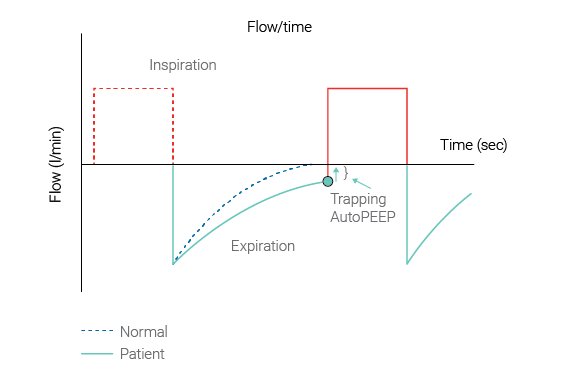
A AutoPEEP é um fenômeno comum em pacientes ventilados mecanicamente com constantes de tempo expiratório longas (RCexp), por exemplo, pacientes com doença pulmonar obstrutiva crônica ou asma aguda grave.

IMPORTANTE: A AutoPEEP resultante não pode ser vista na curva de pressão das vias aéreas exibida na tela do respirador durante o fornecimento normal da respiração.

(Figura 1 abaixo: Source Garcia Vicente et al. (García Vicente E, Sandoval Almengor JC, Díaz Caballero LA, Salgado Campo JC. Ventilación mecánica invasiva en EPOC y asma [Invasive mechanical ventilation in COPD and asthma]. Med Intensiva. 2011;35(5):288-298. doi:10.1016/j.medin.2010.11.0042))

Gráfico de tempo/fluxo exibindo AutoPEEP e retenção de ar
Figura 1: AutoPEEP e retenção de ar
Gráfico de tempo/fluxo exibindo AutoPEEP e retenção de ar
Figura 1: AutoPEEP e retenção de ar

Efeito da AutoPEEP

A AutoPEEP predispõe o paciente ao aumento do trabalho respiratório, barotrauma, instabilidade hemodinâmica e dificuldade de acionar o respirador. O não reconhecimento das consequências hemodinâmicas da AutoPEEP pode levar à restrição inadequada de fluidos ou ao tratamento vasopressor desnecessário. A AutoPEEP pode interferir no desmame da ventilação mecânica.

Os prestadores de cuidados devem monitorar se a AutoPEEP ocorre durante a ventilação e definir os parâmetros de controle da ventilação de acordo para evitar as consequências negativas da AutoPEEP.

Medindo AutoPEEP

Todos os respiradores da Hamilton Medical dispõem da capacidade exclusiva de exibir a AutoPEEP como um parâmetro de monitorização com base em cada ciclo respiratório. É calculado aplicando o MMQ a cada ciclo respiratório (Iotti GA, Braschi A, Brunner JX, et al. Respiratory mechanics by least squares fitting in mechanically ventilated patients: applications during paralysis and during pressure support ventilation. Intensive Care Med. 1995;21(5):406-413. doi:10.1007/BF017074093). No entanto, em circunstâncias especiais, por exemplo, quando ocorre uma hiperinsuflação dinâmica grave, a AutoPEEP calculada pelo MMQ pode distorcer a AutoPEEP real. Nesses casos, é possível obtê-la realizando uma manobra de pausa expiratória.

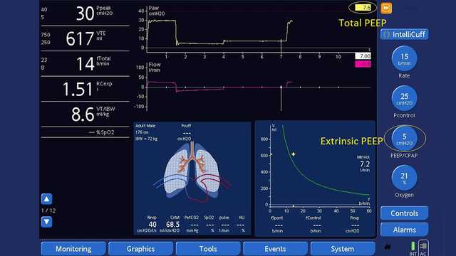
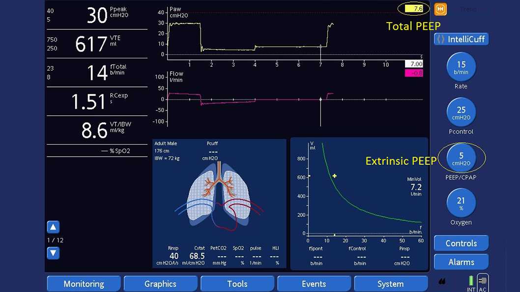
Medindo a PEEP total com uma manobra de pausa expiratória (ver Figura 2 abaixo):

Assegure que a forma de onda Paw é exibida.

  1. Abra a janela Pausa.
  2. Aguarde até que o gráfico da forma de onda Paw reinicie a partir do lado esquerdo.
  3. Aguarde a próxima inspiração.
  4. De seguida, selecione Pausa Exp. Aguarde entre 3 a 5 segundos e, em seguida, selecione Pausa Exp ou pressione o botão P&G novamente para desativar a manobra de pausa e feche a janela.
  5. Após a manobra, a janela Pausa fecha e a função Congelar é ativada automaticamente.
  6. Meça a PEEP total examinando os pontos após o fluxo alcançar zero na curva de pressão com o cursor.
  7. Calcule a AutoPEEP subtraindo a PEEP extrínseca da PEEP total.

Cálculos

AutoPEEP '= PEEP total - PEEP extrínseca = PEEP intrínseca
PEEP '= PEEP extrínseca e está pré-selecionada
PEEP total '= PEEP intrínseca + PEEP extrínseca
Captura de tela da tela do respirador exibindo a manobra de pausa expiratória
Figura 2: Medição da PEEP total usando uma manobra de pausa expiratória - PEEP total de 7,6 cmH2O - PEEP extrínseca de 5 cmH2O = AutoPEEP de 2,6 cmH2O
Captura de tela da tela do respirador exibindo a manobra de pausa expiratória
Figura 2: Medição da PEEP total usando uma manobra de pausa expiratória - PEEP total de 7,6 cmH2O - PEEP extrínseca de 5 cmH2O = AutoPEEP de 2,6 cmH2O

Evitar a retenção de ar

Se a AutoPEEP ocorrer acidentalmente, os prestadores de cuidados devem considerar a adaptação dos parâmetros de controle para evitar a retenção de ar, aumentando o tempo de exalação. O uso de tubos endotraqueais de grande diâmetro, broncodilatadores, tempos inspiratórios curtos, tempos expiratórios longos, frequências respiratórias inferiores e o uso de sedativos podem ser necessários para evitar a hiperinsuflação dinâmica causada pela retenção de ar.

Todos os respiradores Hamilton Medical possuem o modo de ventilação inteligente, Ventilação de Suporte Adaptativo (ASV®). O ASV inclui automaticamente estratégias de proteção pulmonar para minimizar complicações da AutoPEEP.

Dispositivos relevantes: HAMILTON-G5/S1 (software v2.8x e posterior); HAMILTON-C3 (software v2.0.x e posterior), HAMILTON-C6 (software v1.1.x e posterior)

Ver a citação completa de (Iotti, G., & Braschi, A. (1999). Measurements of respiratory mechanics during mechanical ventilation. Rhäzüns, Switzerland: Hamilton Medical Scientific Library.1) abaixo.

Measurements of respiratory mechanics during mechanical ventilation

Iotti, G., & Braschi, A. (1999). Measurements of respiratory mechanics during mechanical ventilation. Rhäzüns, Switzerland: Hamilton Medical Scientific Library.

Invasive mechanical ventilation in COPD and asthma.

García Vicente E, Sandoval Almengor JC, Díaz Caballero LA, Salgado Campo JC. Ventilación mecánica invasiva en EPOC y asma [Invasive mechanical ventilation in COPD and asthma]. Med Intensiva. 2011;35(5):288-298. doi:10.1016/j.medin.2010.11.004

COPD and asthmatic patients use a substantial proportion of mechanical ventilation in the ICU, and their overall mortality with ventilatory support can be significant. From the pathophysiological standpoint, they have increased airway resistance, pulmonary hyperinflation, and high pulmonary dead space, leading to increased work of breathing. If ventilatory demand exceeds work output of the respiratory muscles, acute respiratory failure follows. The main goal of mechanical ventilation in this kind of patients is to improve pulmonary gas exchange and to allow for sufficient rest of compromised respiratory muscles to recover from the fatigued state. The current evidence supports the use of noninvasive positive-pressure ventilation for these patients (especially in COPD), but invasive ventilation also is required frequently in patients who have more severe disease. The physician must be cautious to avoid complications related to mechanical ventilation during ventilatory support. One major cause of the morbidity and mortality arising during mechanical ventilation in these patients is excessive dynamic pulmonary hyperinflation (DH) with intrinsic positive end-expiratory pressure (intrinsic PEEP or auto-PEEP). The purpose of this article is to provide a concise update of the most relevant aspects for the optimal ventilatory management in these patients.

Respiratory mechanics by least squares fitting in mechanically ventilated patients: applications during paralysis and during pressure support ventilation.

Iotti GA, Braschi A, Brunner JX, et al. Respiratory mechanics by least squares fitting in mechanically ventilated patients: applications during paralysis and during pressure support ventilation. Intensive Care Med. 1995;21(5):406-413. doi:10.1007/BF01707409

OBJECTIVE To evaluate a least squares fitting technique for the purpose of measuring total respiratory compliance (Crs) and resistance (Rrs) in patients submitted to partial ventilatory support, without the need for esophageal pressure measurement. DESIGN Prospective, randomized study. SETTING A general ICU of a University Hospital. PATIENTS 11 patients in acute respiratory failure, intubated and assisted by pressure support ventilation (PSV). INTERVENTIONS Patients were ventilated at 4 different levels of pressure support. At the end of the study, they were paralyzed for diagnostic reasons and submitted to volume controlled ventilation (CMV). MEASUREMENTS AND RESULTS A least squares fitting (LSF) method was applied to measure Crs and Rrs at different levels of pressure support as well as in CMV. Crs and Rrs calculated by the LSF method were compared to reference values which were obtained in PSV by measurement of esophageal pressure, and in CMV by the application of the constant flow, end-inspiratory occlusion method. Inspiratory activity was measured by P0.1. In CMV, Crs and Rrs measured by the LSF method are close to quasistatic compliance (-1.5 +/- 1.5 ml/cmH2O) and to the mean value of minimum and maximum end-inspiratory resistance (+0.9 +/- 2.5 cmH2O/(l/s)). Applied during PSV, the LSF method leads to gross underestimation of Rrs (-10.4 +/- 2.3 cmH2O/(l/s)) and overestimation of Crs (+35.2 +/- 33 ml/cmH2O) whenever the set pressure support level is low and the activity of the respiratory muscles is high (P0.1 was 4.6 +/- 3.1 cmH2O). However, satisfactory estimations of Crs and Rrs by the LSF method were obtained at increased pressure support levels, resulting in a mean error of -0.4 +/- 6 ml/cmH2O and -2.8 +/- 1.5 cmH2O/(l/s), respectively. This condition was coincident with a P0.1 of 1.6 +/- 0.7 cmH2O. CONCLUSION The LSF method allows non-invasive evaluation of respiratory mechanics during PSV, provided that a near-relaxation condition is obtained by means of an adequately increased pressure support level. The measurement of P0.1 may be helpful for titrating the pressure support in order to obtain the condition of near-relaxation.