Мы обнаружили, что вы посещаете наш сайт из страны Соединенные Штаты.
Для вашей страны (Соединенные Штаты) доступна отдельная версия веб-сайта.

 Технологии

Волюметрическая капнография Высокотехнологичное измерение концентрации CO2

Изображение лупы

Дополнительные сведения. Волюметрический мониторинг концентрации CO2

Фазы волюметрической капнограммы, форма и морфология кривой, а также измерения, основанные на ее расчетах, могут дать следующую важную информацию:

  • об эффективности вентиляции и перфузии;
  • о физиологической объемной части мертвого пространства;
  • о скорости метаболизма у пациента (Jaffe MB. Using the features of the time and volumetric capnogram for classification and prediction. J Clin Monit Comput. 2017;31(1):19-41. doi:10.1007/s10877-016-9830-z1​)
Датчик CO2 CAPNOSTAT-5 для основного потока

Мощный инструмент. Датчик CO2

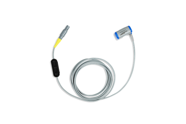
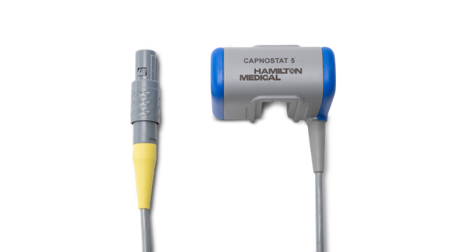
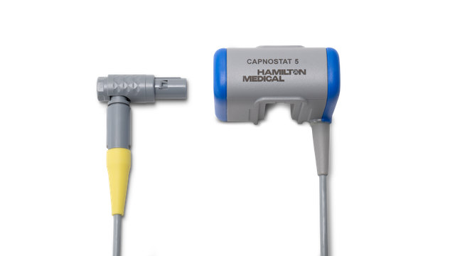
В наших аппаратах ИВЛ концентрация CO2 измеряется с помощью СО2-датчика для основного потока CAPNOSTAT-5, который находится рядом с дыхательными путями пациента.

Датчик CAPNOSTAT-5 обеспечивает точное измерение концентрации углекислого газа в конце выдоха (PetCO2), а также дает точную капнограмму при частоте дыхания до 150 дыхательных движений в минуту.

Графическое представление статистических данных: анализ данных датчика CO2

Маленький датчик, большой объем данных Информация, которую передает датчик

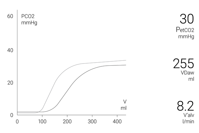
В окне волюметрической капнограммы на экране отображается точная количественная информация в виде комбинации данных проксимального потока и уровня CO2, таких как:

  • текущая кривая волюметрической капнограммы;
  • эталонная кривая волюметрической капнограммы;
  • кнопка эталонной кривой со временем и датой референсной петли;
  • наиболее актуальные значения CO2 для каждого вдоха.

Для более полного анализа состояния пациента в аппаратах ИВЛ предусмотрена функция определения трендов за 72 часа (в аппаратах ИВЛ HAMILTON-G5/S1 – за 96 часов) для следующих параметров:

  • «PetCO2»;
  • «V'CO2»;
  • «FetCO2»;
  • «VeCO2»;
  • «ViCO2»;
  • «Vtальв»;
  • «V'альв»;
  • «VDдп»;
  • «VD/Vt»;
  • «VDдп/VTE»;
  • «НарастCO2»

Для удобства пользователя в аппаратах ИВЛ Hamilton Medical все значения, связанные с CO2, отображаются в окне «Мониторинг CO2».

  • Парциальная концентрация CO2 в конце выдоха: FetCO2 (%) 
  • Давление CO2 в конце выдоха: PetCO2 (ммРт) 
  • Подъем альвеолярного плато на кривой значений «PetCO2», указывающий на показатели объема/потока в легких: нарастCO2 (%CO2/л)
  • Альвеолярный дыхательный объем: Vtальв (мл) 
  • Альвеолярная минутная вентиляция: V'альв (л/мин) 
  • Выведение CO2: V’CO2 (мл/мин) 
  • Мертвое пространство дыхательных путей: VDдп (мл)
  • Объемная часть мертвого пространства на входе в дыхательные пути: VDдп/VTE (%) 
  • Объем выдыхаемого CO2: VeCO2 (мл) 
  • Объем вдыхаемого CO2: ViCO2 (мл)
Электронная книга о волюметрической капнографии

Бесплатная электронная книга

Полезно знать! Все о волюметрической капнографии

Узнайте, как читать волюметрические капнограммы, каковы их преимущества и где применять полученные данные. Книга также содержит тест для самопроверки.

Графическое представление статистических данных: www.hamilton-medical.com/capnography

Каковы преимущества? Немного доказательств

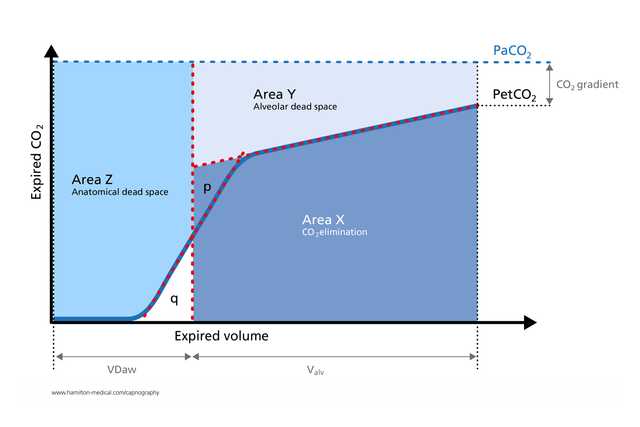
  • Волюметрическая капнограмма успешно используется для измерения анатомического мертвого пространства, перфузии легочных капилляров и эффективности вентиляции (Romero PV, Lucangelo U, Lopez Aguilar J, Fernandez R, Blanch L. Physiologically based indices of volumetric capnography in patients receiving mechanical ventilation. Eur Respir J. 1997;10(6):1309-1315. doi:10.1183/09031936.97.100613092​)

  • Расчеты, полученные с помощью волюметрической капнографии, используются для выявления эмболии сосудов легких у постели больного (Blanch L, Romero PV, Lucangelo U. Volumetric capnography in the mechanically ventilated patient. Minerva Anestesiol. 2006;72(6):577-585. 3​)

  • Исследование показало, что при волюметрической капнографии результаты измерения соотношения физиологической объемной части мертвого пространства и дыхательного объема у пациентов с ARDS, находящихся на искусственной вентиляции легких, отличаются такой же точностью, как и результаты, полученные методом метаболического мониторинга (Kallet RH, Daniel BM, Garcia O, Matthay MA. Accuracy of physiologic dead space measurements in patients with acute respiratory distress syndrome using volumetric capnography: comparison with the metabolic monitor method. Respir Care. 2005;50(4):462-467. 4​)

  • Экспираторная капнограмма – это быстрое неинвазивное измерение, не зависящее от дыхательного усилия, которое помогает выявить выраженный бронхоспазм у взрослых пациентов с астмой (Yaron M, Padyk P, Hutsinpiller M, Cairns CB. Utility of the expiratory capnogram in the assessment of bronchospasm. Ann Emerg Med. 1996;28(4):403-407. doi:10.1016/s0196-0644(96)70005-75​)

  • Волюметрическая капнография дает ценную информацию о физиологии коллапса легких и рекрутмента неинвазивным способом в реальном времени и таким образом позволяет контролировать цикличное выполнение маневров рекрутмента при лечении пациента (Tusman G, Suarez-Sipmann F, Böhm SH, et al. Monitoring dead space during recruitment and PEEP titration in an experimental model. Intensive Care Med. 2006;32(11):1863-1871. doi:10.1007/s00134-006-0371-76​)

Изображение студентки с сертификатом в руке

Полезно знать! Материалы курса обучения по использованию волюметрической капнографии

Принадлежности и расходные материалы

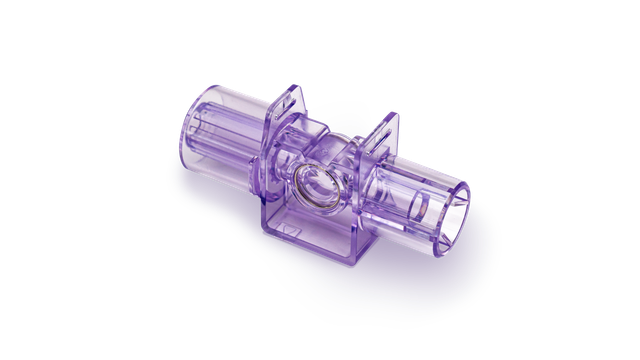
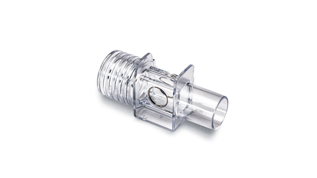
Компания Hamilton Medical предлагает оригинальные расходные материалы для всех групп пациентов: взрослых, детей и младенцев. Доступны продукты для многократного или разового использования, (выбор зависит от политики вашего учреждения).

Доступность

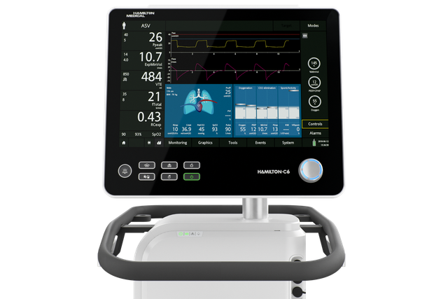
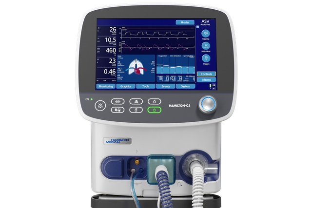
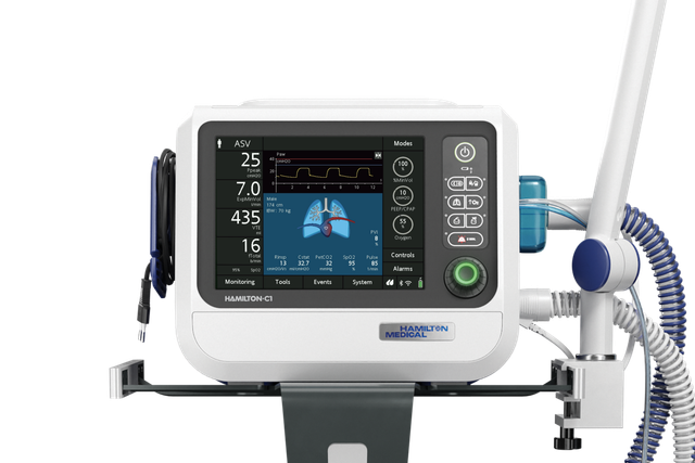
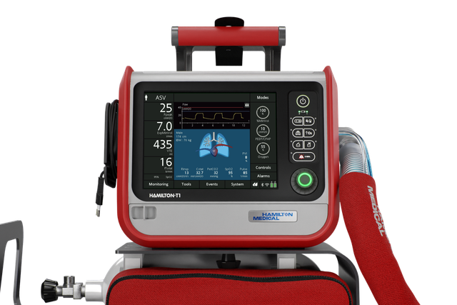
Волюметрическая капнография доступна как опция в аппаратах ИВЛ HAMILTON-C6, HAMILTON-G5, HAMILTON-C3 и HAMILTON-C1/T1, а также как стандартная функция в аппарате HAMILTON-S1.

Using the features of the time and volumetric capnogram for classification and prediction.

Jaffe MB. Using the features of the time and volumetric capnogram for classification and prediction. J Clin Monit Comput. 2017;31(1):19-41. doi:10.1007/s10877-016-9830-z

Quantitative features derived from the time-based and volumetric capnogram such as respiratory rate, end-tidal PCO2, dead space, carbon dioxide production, and qualitative features such as the shape of capnogram are clinical metrics recognized as important for assessing respiratory function. Researchers are increasingly exploring these and other known physiologically relevant quantitative features, as well as new features derived from the time and volumetric capnogram or transformations of these waveforms, for: (a) real-time waveform classification/anomaly detection, (b) classification of a candidate capnogram into one of several disease classes, (c) estimation of the value of an inaccessible or invasively determined physiologic parameter, (d) prediction of the presence or absence of disease condition, (e) guiding the administration of therapy, and (f) prediction of the likely future morbidity or mortality of a patient with a presenting condition. The work to date with respect to these applications will be reviewed, the underlying algorithms and performance highlighted, and opportunities for the future noted.

Physiologically based indices of volumetric capnography in patients receiving mechanical ventilation.

Romero PV, Lucangelo U, Lopez Aguilar J, Fernandez R, Blanch L. Physiologically based indices of volumetric capnography in patients receiving mechanical ventilation. Eur Respir J. 1997;10(6):1309-1315. doi:10.1183/09031936.97.10061309

Several indices of ventilatory heterogeneity can be identified from the expiratory CO2 partial pressure or CO2 elimination versus volume curves. The aims of this study were: 1) to analyse several computerizable indices of volumetric capnography in order to detect ventilatory disturbances; and 2) to establish the relationship between those indices and respiratory system mechanics in subjects with normal lungs and in patients with acute respiratory distress syndrome (ARDS), both receiving mechanical ventilation. We studied six normal subjects and five patients with early ARDS mechanically ventilated at three levels of tidal volume (VT). Respiratory system mechanics were assessed by end-expiratory and end-inspiratory occlusion methods, respectively. We determined Phase III slopes, Fletcher's efficiency index, Bohr's dead space (VD,Bohr/VT), and the ratio of alveolar ejection volume to tidal volume (VAE/VT) from expiratory capnograms, as a function of expired volume. Differences between normal subjects and ARDS patients were significant both for capnographic and mechanical parameters. Changes in VT significantly altered capnographic indices in normal subjects, but failed to change ventilatory mechanics and VAE/VT in ARDS patients. After adjusting for breathing pattern, VAE/VT exhibited the best correlation with the mechanical parameters. In conclusion, volumetric capnography, and, specifically, the ratio of alveolar ejection volume to tidal volume allows evaluation and monitoring of ventilatory disturbances in patients with adult respiratory distress syndrome.

Volumetric capnography in the mechanically ventilated patient.

Blanch L, Romero PV, Lucangelo U. Volumetric capnography in the mechanically ventilated patient. Minerva Anestesiol. 2006;72(6):577-585.

Expiratory capnogram provides qualitative information on the waveform patterns associated with mechanical ventilation and quantitative estimation of expired CO2. Volumetric capnography simultaneously measures expired CO2 and tidal volume and allows identification of CO2 from 3 sequential lung compartments: apparatus and anatomic dead space, from progressive emptying of alveoli and alveolar gas. Lung heterogeneity creates regional differences in CO2 concentration and sequential emptying contributes to the rise of the alveolar plateau and to the steeper the expired CO2 slope. The concept of dead space accounts for those lung areas that are ventilated but not perfused. In patients with sudden pulmonary vascular occlusion due to pulmonary embolism, the resultant high V/Q mismatch produces an increase in alveolar dead space. Calculations derived from volumetric capnography are useful to suspect pulmonary embolism at the bedside. Alveolar dead space is large in acute lung injury and when the effect of positive end-expiratory pressure (PEEP) is to recruit collapsed lung units resulting in an improvement of oxygenation, alveolar dead space may decrease, whereas PEEP-induced overdistension tends to increase alveolar dead space. Finally, measurement of physiologic dead space and alveolar ejection volume at admission or the trend during the first 48 hours of mechanical ventilation might provide useful information on outcome of critically ill patients with acute lung injury or acute respiratory distress syndrome.

Accuracy of physiologic dead space measurements in patients with acute respiratory distress syndrome using volumetric capnography: comparison with the metabolic monitor method.

Kallet RH, Daniel BM, Garcia O, Matthay MA. Accuracy of physiologic dead space measurements in patients with acute respiratory distress syndrome using volumetric capnography: comparison with the metabolic monitor method. Respir Care. 2005;50(4):462-467.


BACKGROUND

Volumetric capnography is an alternative method of measuring expired carbon dioxide partial pressure (P(eCO2)) and physiologic dead-space-to-tidal-volume ratio (V(D)/V(T)) during mechanical ventilation. In this method, P(eCO2) is measured at the Y-adapter of the ventilator circuit, thus eliminating the effects of compression volume contamination and the need to apply a correction factor. We investigated the accuracy of volumetric capnography in measuring V(D)/V(T), compared to both uncorrected and corrected measurements, using a metabolic monitor in patients with acute respiratory distress syndrome (ARDS).

METHODS

There were 90 measurements of V(D)/V(T) made in 23 patients with ARDS. The P(eCO2) was measured during a 5-min expired-gas collection period with a Delta-trac metabolic monitor, and was corrected for compression volume contamination using a standard formula. Simultaneous measurements of P(eCO2) and V(D)/V(T) were obtained using volumetric capnography.

RESULTS

V(D)/V(T) measured by volumetric capnography was strongly correlated with both the uncorrected (r2 = 0.93, p < 0.0001) and corrected (r2 = 0.89, p < 0.0001) measurements of V(D)/V(T) made using the metabolic monitor technique. Measurements of V(D)/V(T) made with volumetric capnography had a bias of 0.02 and a precision of 0.05 when compared to the V(D)/V(T) corrected for estimated compression volume contamination.

CONCLUSION

Volumetric capnography measurements of V(D)/V(T) in mechanically-ventilated patients with ARDS are as accurate as those obtained by metabolic monitor technique. .

Utility of the expiratory capnogram in the assessment of bronchospasm.

Yaron M, Padyk P, Hutsinpiller M, Cairns CB. Utility of the expiratory capnogram in the assessment of bronchospasm. Ann Emerg Med. 1996;28(4):403-407. doi:10.1016/s0196-0644(96)70005-7


STUDY OBJECTIVE

To determine whether the plateau phase of the expiratory capnogram (dco2/dt) can detect bronchospasm in adult asthma patients in the emergency department and to assess the correlation between dco2/dt and the peak expiratory flow rate (PEFR) in spontaneously breathing patients with asthma and in normal, healthy volunteers.

METHODS

We carried out a prospective, blinded study in a university hospital ED. Twenty adults (12 women) with acute asthma and 28 normal adult volunteers (15 women) breathed through the sampling probe of an end-tidal CO2 monitor, and the expired CO2 waveform was recorded. The dco2/dt of the plateau (alveolar) phase for five consecutive regular expirations was measured and a mean value calculated for each patient. The best of three PEFRs was determined. The PEFR and dco2/dt were also recorded after treatment of the asthmatic patients with inhaled beta-agonists.

RESULTS

The mean +/- SD PEFR of the asthmatic subjects was 274 +/- 96 L/minute (57% of the predicted value), whereas that of the normal volunteers was 527 +/- 96 L/minute (103% of the predicted value) (P < .001). The mean dco2/dt of the asthmatic subjects (.26 +/- .06) was significantly steeper than that of the normal volunteers (.13 +/- .06) (P < .001). The dco2/dt was correlated with PEFR (r = .84, P < .001). In 18 asthmatic subjects the pretreatment and posttreatment percent predicted PEFRS were 58% +/- 17% and 74% +/- 17%, respectively (P < .001), whereas the dco2/dt values were .27 +/- .05 and .19 +/- .07, respectively (P < .005).

CONCLUSION

The dco2/dt is an effort-independent, rapid noninvasive measure that indicates significant bronchospasm in ED adult patients with asthma. The dco2/dt value is correlated with PEFR, an effort-dependent measure of airway obstruction. The change in dco2/dt with inhaled beta-agonists may be useful in monitoring the therapy of acute asthma.

Monitoring dead space during recruitment and PEEP titration in an experimental model.

Tusman G, Suarez-Sipmann F, Böhm SH, et al. Monitoring dead space during recruitment and PEEP titration in an experimental model. Intensive Care Med. 2006;32(11):1863-1871. doi:10.1007/s00134-006-0371-7


OBJECTIVE

To test the usefulness of dead space for determining open-lung PEEP, the lowest PEEP that prevents lung collapse after a lung recruitment maneuver.

DESIGN

Prospective animal study.

SETTING

Department of Clinical Physiology, University of Uppsala, Sweden.

SUBJECTS

Eight lung-lavaged pigs.

INTERVENTIONS

Animals were ventilated using constant flow mode with VT of 6ml/kg, respiratory rate of 30bpm, inspiratory-to-expiratory ratio of 1:2, and FiO(2) of 1. Baseline measurements were performed at 6cmH(2)O of PEEP. PEEP was increased in steps of 6cmH(2)O from 6 to 24cmH(2)O. Recruitment maneuver was achieved within 2min at pressure levels of 60/30cmH(2)O for Peak/PEEP. PEEP was decreased from 24 to 6cmH(2)O in steps of 2cmH(2)O and then to 0cmH(2)O. Each PEEP step was maintained for 10min.

MEASUREMENTS AND RESULTS

Alveolar dead space (VD(alv)), the ratio of alveolar dead space to alveolar tidal volume (VD(alv)/VT(alv)), and the arterial to end-tidal PCO(2) difference (Pa-ET: CO(2)) showed a good correlation with PaO(2), normally aerated areas, and non-aerated CT areas in all animals (minimum-maximum r(2)=0.83-0.99; p<0.01). Lung collapse (non-aerated tissue>5%) started at 12[Symbol: see text]cmH(2)O PEEP; hence, open-lung PEEP was established at 14cmH(2)O. The receiver operating characteristics curve demonstrated a high specificity and sensitivity of VD(alv) (0.89 and 0.90), VD(alv)/VT(alv) (0.82 and 1.00), and Pa-ET: CO(2) (0.93 and 0.95) for detecting lung collapse.

CONCLUSIONS

Monitoring of dead space was useful for detecting lung collapse and for establishing open-lung PEEP after a recruitment maneuver.