Назад

Рекомендации по правильному измерению пищеводного давления

Статья

Автор: Жан-Мишель Арналь, старший реаниматолог, больница Hopital Sainte Musse, Тулон, Франция

Дата: 19.10.2018

Last change: 09.09.2020

Formatting

Недавнее физиологическое исследование показало, что пищеводное давление определяет плевральное давление в середине грудной полости при всех уровнях PEEP. Таким образом, абсолютное измерение пищеводного давления помогает при установке значения PEEP и мониторинге транспульмонального давления.

Рекомендации по правильному измерению пищеводного давления

Первоначальное размещение и наполнение воздухом

Так как же следует правильно измерять пищеводное давление?

Пищеводный баллон необходимо правильно расположить и наполнить воздухом, а затем следует проверить его размещение.

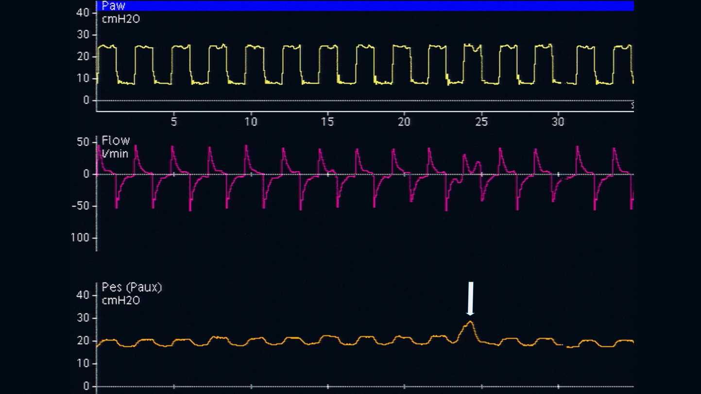
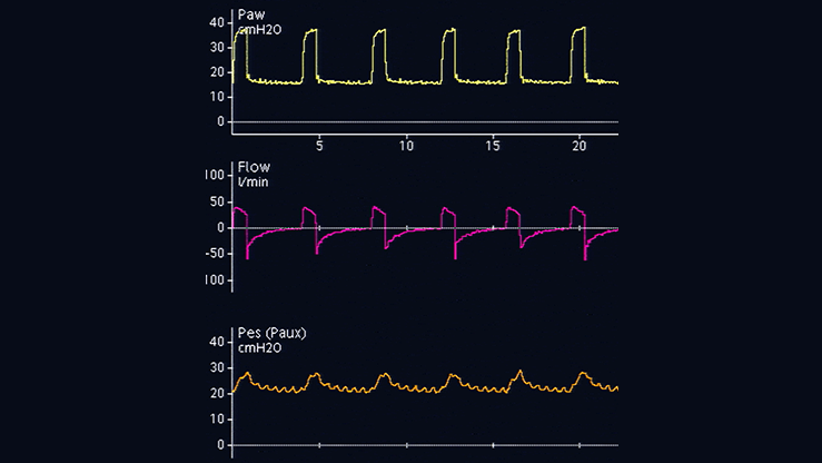
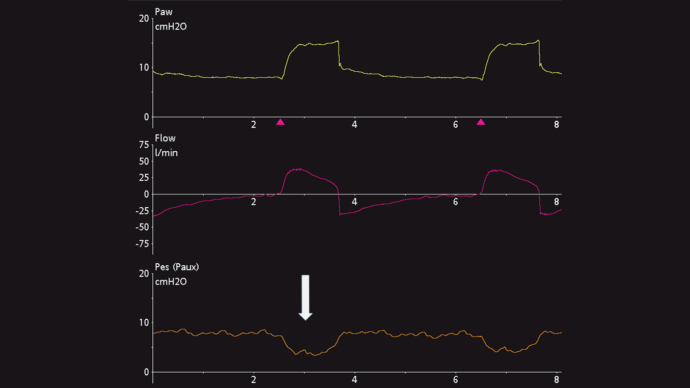
Оптимальное положение пищеводного баллона – нижняя треть пищевода, расстояние от ноздрей – 35–45 см. Пациент должен находиться в положении полулежа. Пустой баллон сначала вводится в желудок, расположенный примерно в 50–60 см от ноздрей. Баллон наполняют воздухом до стандартного объема (1 мл для катетера CooperSurgical и 4 мл для катетера Nutrivent). Во время вдоха внутрижелудочное давление имеет положительное отклонение как у пассивных, так и у спонтанно дышащих пациентов. Положение желудка проверяют путем аккуратного нажатия руками на эпигастральную область, вследствие чего внутрижелудочное давление сразу повышается (см. рисунок 1).

Снимок экрана с кривой, на которой показано увеличение значения Pes
Рисунок 1
Снимок экрана с кривой, на которой показано увеличение значения Pes
Рисунок 1

Извлечение с наполненным баллоном

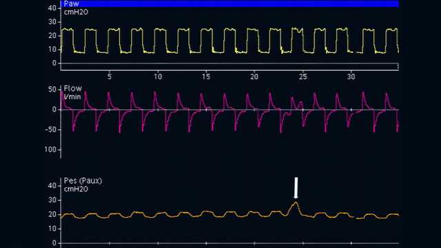
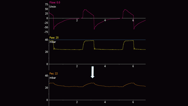
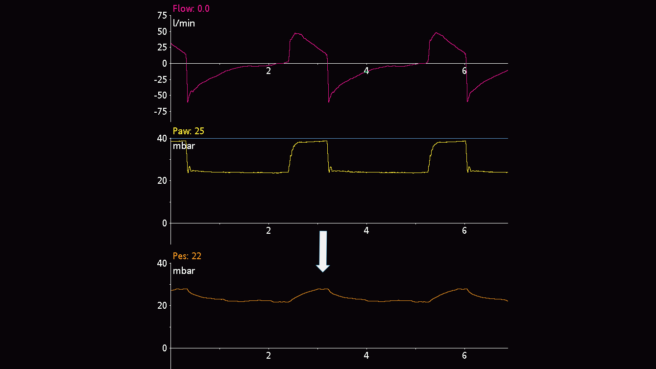
Затем пищеводный катетер осторожно извлекают, пока баллон все еще наполнен, чтобы расположить баллон в нижней трети пищевода. При переходе от внутрижелудочного давления (см. рисунок 2) к пищеводному (см. рисунок 3) давления исходная линия кривой давления меняется, и отображает сердечные осцилляции.

Исходная кривая внутрижелудочного давления
Рисунок 2. Внутрижелудочное давление
Исходная кривая внутрижелудочного давления
Рисунок 2. Внутрижелудочное давление
Исходная кривая пищеводного давления
Рисунок 3. Пищеводное давление
Исходная кривая пищеводного давления
Рисунок 3. Пищеводное давление

Отклонения пищеводного давления

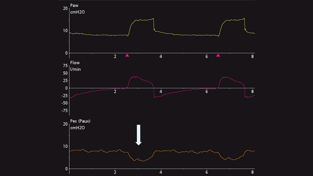
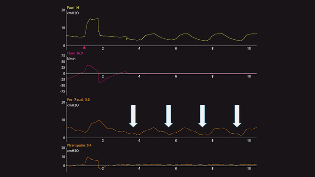
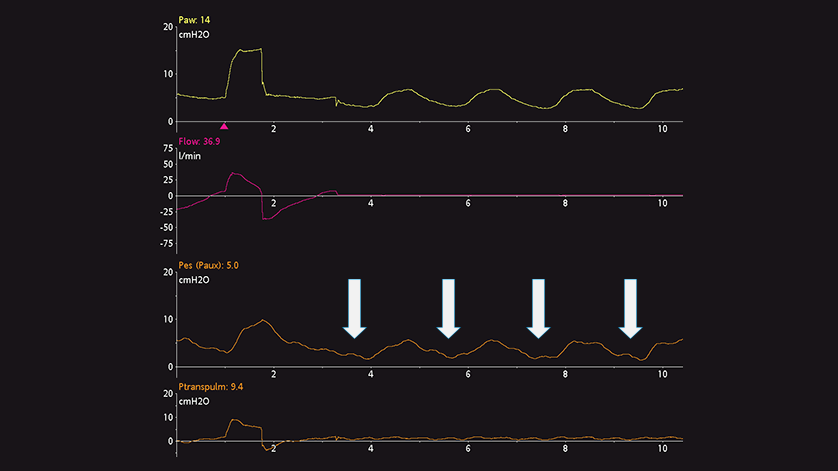
Отклонения пищеводного давления положительны во время вдоха у пассивных пациентов (см. рисунок 4), но отрицательны у пациентов со спонтанным дыханием (см. рисунок 5). Если сердечные осцилляции искажают сигнал пищеводного давления, можно извлечь катетер еще на 2–5 см.
 

Кривая отклонений значения Pes у пассивного пациента
Рисунок 4. Пассивный пациент
Кривая отклонений значения Pes у пассивного пациента
Рисунок 4. Пассивный пациент
Кривая отклонений значения Pes у спонтанно дышащего пациента
Рисунок 5. Спонтанно дышащий пациент
Кривая отклонений значения Pes у спонтанно дышащего пациента
Рисунок 5. Спонтанно дышащий пациент

Наполнение баллона воздухом

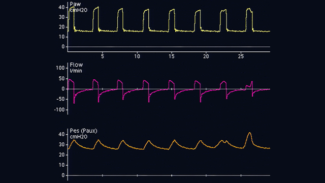
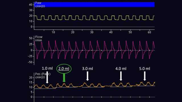
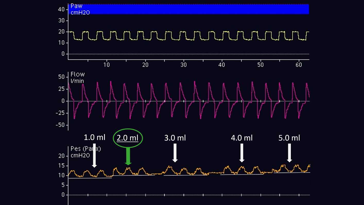
Объем воздуха для правильного наполнения баллона следует титровать индивидуально. Это возможно только у пассивных пациентов. Согласно методу, предложенному Моджоли и др. в 2016 г., баллон наполняют на 0,5–3 мл с постепенным шагом 0,5 мл для катетера Cooper Surgical и на1–8 мл с шагом 1 мл для катетера Nutrivent (см. рисунок 6). Во время постепенного наполнения баллона исходное пищеводное давление увеличивается, а величина отклонения пищеводного давления изменяется. Правильный объем наполнения определяется по наибольшему отклонению пищеводного давления. Если при двух разных объемах наполнения величина отклонения пищеводного давления одинакова, выбирается наименьший объем наполнения.

Кривая во время наполнения баллона
Рисунок 6. Наполнение баллона с помощью катетера Nutrivent
Кривая во время наполнения баллона
Рисунок 6. Наполнение баллона с помощью катетера Nutrivent

Контрольная проверка

После правильного расположения баллона в пищеводе и его наполнения проводится контрольная проверка наличия окклюзии. При проверке дыхательные пути перекрывают в конце выдоха, чтобы изменить давление в них, а затем проверяют, изменилось ли пищеводное давление на ту же величину.

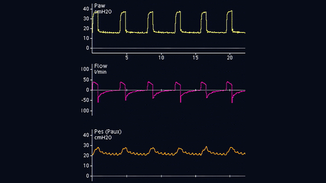
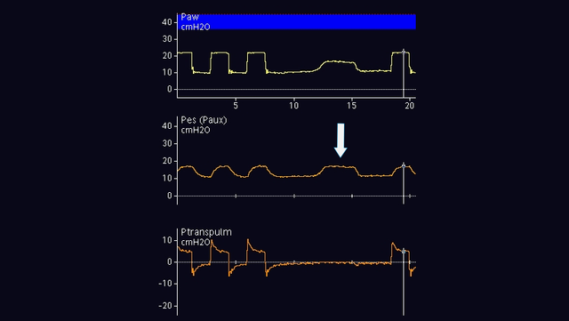
У пассивных пациентов можно выполнить окклюзию в конце выдоха. Когда клапан выдоха закрыт, сожмите грудную клетку руками с обеих сторон, чтобы увидеть положительное отклонение давления в дыхательных путях и пищеводе. Величина повышения давления в дыхательных путях и пищеводе должна быть одинаковой. Другими словами, транспульмональное давление не должно изменяться (см. рисунок 7).

У активных пациентов проверка наличия динамической окклюзии также включает окклюзию в конце выдоха. Нет необходимости сжимать грудную клетку руками, так как во время окклюзии пациент будет совершать спонтанное дыхательное усилие. Результатом является отрицательное отклонение давления в дыхательных путях и пищеводе. Величина снижения давления в дыхательных путях и пищеводе должна быть одинаковой, т. е. транспульмональное давление не должно изменяться (см. рисунок 8).

Если требуется постоянно контролировать пищеводное давление, необходимо повторно проверить правильность размещения и объем наполнения баллона.

Полный список цитируемых материалов см. ниже: (Yoshida T, Amato MBP, Grieco DL, et al. Esophageal Manometry and Regional Transpulmonary Pressure in Lung Injury. Am J Respir Crit Care Med. 2018;197(8):1018-1026. doi:10.1164/rccm.201709-1806OC1​, Mojoli F, Iotti GA, Torriglia F, et al. In vivo calibration of esophageal pressure in the mechanically ventilated patient makes measurements reliable. Crit Care. 2016;20:98. Published 2016 Apr 11. doi:10.1186/s13054-016-1278-52​, Baydur A, Behrakis PK, Zin WA, Jaeger M, Milic-Emili J. A simple method for assessing the validity of the esophageal balloon technique. Am Rev Respir Dis. 1982;126(5):788-791. doi:10.1164/arrd.1982.126.5.7883​, Akoumianaki E, Maggiore SM, Valenza F, et al. The application of esophageal pressure measurement in patients with respiratory failure. Am J Respir Crit Care Med. 2014;189(5):520-531. doi:10.1164/rccm.201312-2193CI4​, Mauri T, Yoshida T, Bellani G, et al. Esophageal and transpulmonary pressure in the clinical setting: meaning, usefulness and perspectives. Intensive Care Med. 2016;42(9):1360-1373. doi:10.1007/s00134-016-4400-x5​)

Кривая, показывающая увеличение значений Paw и Pes; значение Ptranspulm не изменилось
Рисунок 7. Проверка наличия окклюзии у пассивного пациента
Кривая, показывающая увеличение значений Paw и Pes; значение Ptranspulm не изменилось
Рисунок 7. Проверка наличия окклюзии у пассивного пациента
Кривая, показывающая снижение значений Paw и Pes; значение Ptranspulm не изменилось
Рисунок 8. Проверка наличия окклюзии у активного пациента
Кривая, показывающая снижение значений Paw и Pes; значение Ptranspulm не изменилось
Рисунок 8. Проверка наличия окклюзии у активного пациента
Электронная книга, которая содержит 13 советов от специалистов

Бесплатная электронная книга

13 советов от специалистов. Измерение пищеводного давления

Клинически проверенные рекомендации касательно того, что следует делать и чего следует избегать при измерении пищеводного давления у пациентов с острым респираторным дистресс-синдромом.

Esophageal Manometry and Regional Transpulmonary Pressure in Lung Injury.

Yoshida T, Amato MBP, Grieco DL, et al. Esophageal Manometry and Regional Transpulmonary Pressure in Lung Injury. Am J Respir Crit Care Med. 2018;197(8):1018-1026. doi:10.1164/rccm.201709-1806OC


RATIONALE

Esophageal manometry is the clinically available method to estimate pleural pressure, thus enabling calculation of transpulmonary pressure (Pl). However, many concerns make it uncertain in which lung region esophageal manometry reflects local Pl.

OBJECTIVES

To determine the accuracy of esophageal pressure (Pes) and in which regions esophageal manometry reflects pleural pressure (Ppl) and Pl; to assess whether lung stress in nondependent regions can be estimated at end-inspiration from Pl.

METHODS

In lung-injured pigs (n = 6) and human cadavers (n = 3), Pes was measured across a range of positive end-expiratory pressure, together with directly measured Ppl in nondependent and dependent pleural regions. All measurements were obtained with minimal nonstressed volumes in the pleural sensors and esophageal balloons. Expiratory and inspiratory Pl was calculated by subtracting local Ppl or Pes from airway pressure; inspiratory Pl was also estimated by subtracting Ppl (calculated from chest wall and respiratory system elastance) from the airway plateau pressure.

MEASUREMENTS AND MAIN RESULTS

In pigs and human cadavers, expiratory and inspiratory Pl using Pes closely reflected values in dependent to middle lung (adjacent to the esophagus). Inspiratory Pl estimated from elastance ratio reflected the directly measured nondependent values.

CONCLUSIONS

These data support the use of esophageal manometry in acute respiratory distress syndrome. Assuming correct calibration, expiratory Pl derived from Pes reflects Pl in dependent to middle lung, where atelectasis usually predominates; inspiratory Pl estimated from elastance ratio may indicate the highest level of lung stress in nondependent "baby" lung, where it is vulnerable to ventilator-induced lung injury.

In vivo calibration of esophageal pressure in the mechanically ventilated patient makes measurements reliable.

Mojoli F, Iotti GA, Torriglia F, et al. In vivo calibration of esophageal pressure in the mechanically ventilated patient makes measurements reliable. Crit Care. 2016;20:98. Published 2016 Apr 11. doi:10.1186/s13054-016-1278-5

In screening programmes it is important to assess a preliminary effectiveness of the screening method as soon as possible in order to forecast survival figures. In March 1981 a controlled single-view mammographic screening trial for breast cancer was started in the south of Stockholm. The population invited for screening mammography consisted of 40,000 women aged 40-64 years, and 20,000 women served as a well-defined control group. The main aim of the trial was to determine whether repeated mammographic screening could reduce the mortality in the study population (SP) compared to the control population (CP). The cumulative number of advanced mammary carcinomas in the screening and the control populations from the first five years of screening have shown a tendency towards more favourable stages in the screened population aged 40-64 years. A breakdown by age suggests an effect in age group 50-59 years, but not yet in age groups 40-49 and 60-64 years. When comparing the rates of stage II+ cancer, an increased number is found in the study group. As the total rate of breast cancer is higher in SP than in CP, there ought to be a concealed group of stage II+ cancers in the CP which makes the comparison biased. A new approach has been designed, where an estimation of the 'hidden' number of stage II+ cancers in CP is added to the clinically detected cases, and in this respect a comparison has shown a decrease in the cumulative number of advanced cancers in the SP in relation to the CP (p less than 0.05).(ABSTRACT TRUNCATED AT 250 WORDS)

A simple method for assessing the validity of the esophageal balloon technique.

Baydur A, Behrakis PK, Zin WA, Jaeger M, Milic-Emili J. A simple method for assessing the validity of the esophageal balloon technique. Am Rev Respir Dis. 1982;126(5):788-791. doi:10.1164/arrd.1982.126.5.788

The validity of the conventional esophageal balloon technique as a measure of pleural pressure was tested in 10 subjects in sitting, supine, and lateral positions by occluding the airways at end-expiration and measuring the ratio of changes in esophageal (delta Pes) and mouth pressure (delta Pm) during the ensuing spontaneous occluded inspiratory efforts. Similar measurements were also made during static Mueller maneuvers. In both tests, delta Pes/delta Pm values were close to unity in sitting and lateral positions, whereas in the supine position, substantial deviations from unity were found in some instances. However, by repositioning the balloon to different levels in the esophagus, even in these instances a locus could be found where the delta Pes/delta Pm ratio was close to unity. No appreciable phase difference between delta Pes and delta Pm was found. We conclude that by positioning the balloon according to the "occlusion test" procedure, valid measurements of pleural pressure can be obtained in all the tested body positions.

The application of esophageal pressure measurement in patients with respiratory failure.

Akoumianaki E, Maggiore SM, Valenza F, et al. The application of esophageal pressure measurement in patients with respiratory failure. Am J Respir Crit Care Med. 2014;189(5):520-531. doi:10.1164/rccm.201312-2193CI

This report summarizes current physiological and technical knowledge on esophageal pressure (Pes) measurements in patients receiving mechanical ventilation. The respiratory changes in Pes are representative of changes in pleural pressure. The difference between airway pressure (Paw) and Pes is a valid estimate of transpulmonary pressure. Pes helps determine what fraction of Paw is applied to overcome lung and chest wall elastance. Pes is usually measured via a catheter with an air-filled thin-walled latex balloon inserted nasally or orally. To validate Pes measurement, a dynamic occlusion test measures the ratio of change in Pes to change in Paw during inspiratory efforts against a closed airway. A ratio close to unity indicates that the system provides a valid measurement. Provided transpulmonary pressure is the lung-distending pressure, and that chest wall elastance may vary among individuals, a physiologically based ventilator strategy should take the transpulmonary pressure into account. For monitoring purposes, clinicians rely mostly on Paw and flow waveforms. However, these measurements may mask profound patient-ventilator asynchrony and do not allow respiratory muscle effort assessment. Pes also permits the measurement of transmural vascular pressures during both passive and active breathing. Pes measurements have enhanced our understanding of the pathophysiology of acute lung injury, patient-ventilator interaction, and weaning failure. The use of Pes for positive end-expiratory pressure titration may help improve oxygenation and compliance. Pes measurements make it feasible to individualize the level of muscle effort during mechanical ventilation and weaning. The time is now right to apply the knowledge obtained with Pes to improve the management of critically ill and ventilator-dependent patients.

Esophageal and transpulmonary pressure in the clinical setting: meaning, usefulness and perspectives.

Mauri T, Yoshida T, Bellani G, et al. Esophageal and transpulmonary pressure in the clinical setting: meaning, usefulness and perspectives. Intensive Care Med. 2016;42(9):1360-1373. doi:10.1007/s00134-016-4400-x


PURPOSE

Esophageal pressure (Pes) is a minimally invasive advanced respiratory monitoring method with the potential to guide management of ventilation support and enhance specific diagnoses in acute respiratory failure patients. To date, the use of Pes in the clinical setting is limited, and it is often seen as a research tool only.

METHODS

This is a review of the relevant technical, physiological and clinical details that support the clinical utility of Pes.

RESULTS

After appropriately positioning of the esophageal balloon, Pes monitoring allows titration of controlled and assisted mechanical ventilation to achieve personalized protective settings and the desired level of patient effort from the acute phase through to weaning. Moreover, Pes monitoring permits accurate measurement of transmural vascular pressure and intrinsic positive end-expiratory pressure and facilitates detection of patient-ventilator asynchrony, thereby supporting specific diagnoses and interventions. Finally, some Pes-derived measures may also be obtained by monitoring electrical activity of the diaphragm.

CONCLUSIONS

Pes monitoring provides unique bedside measures for a better understanding of the pathophysiology of acute respiratory failure patients. Including Pes monitoring in the intensivist's clinical armamentarium may enhance treatment to improve clinical outcomes.