Back

Basics of volumetric capnography - Part 2: Single breath CO2 analysis

Article

Author: Karjaghli Munir, Respiratory Therapist, Hamilton Medical Clinical Application Specialist; Matthias Himmelstoss, ICU Nurse, MSc Physics, Product Manager

Date of first publication: 16.11.2023

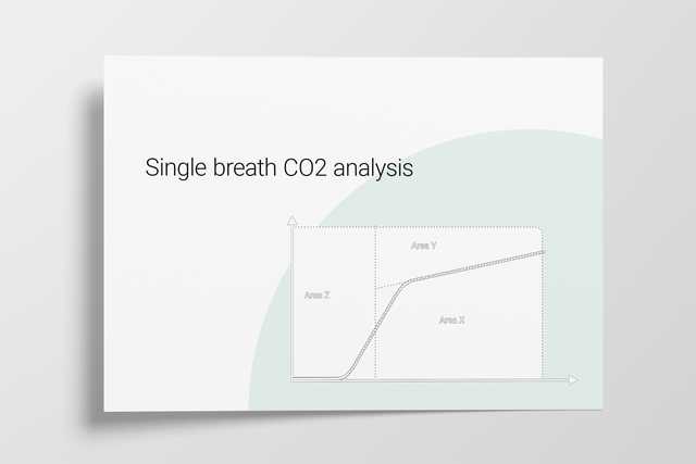
Anatomical dead space, alveolar dead space, and CO2 elimination: Learn how they can give you an insight into the patient's lung condition.

Basics of volumetric capnography - Part 2: Single breath CO2 analysis

Single breath CO2 analysis: insight into the patient‘s lung condition

The volumetric capnogram can also be divided into three areas:

  • Area X - CO2 elimination
  • Area Y - Alveolar dead space
  • Area Z - Anatomical dead space

The size of the areas, as well as the form of the curve, can give you more insight into the patient‘s lung condition regarding:

  • Dead space fraction – VDaw /VTE
  • Alveolar minute ventilation – V‘alv

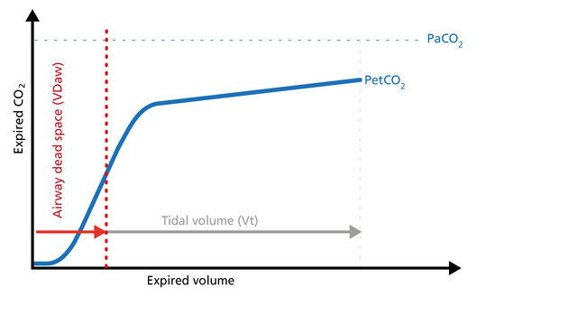
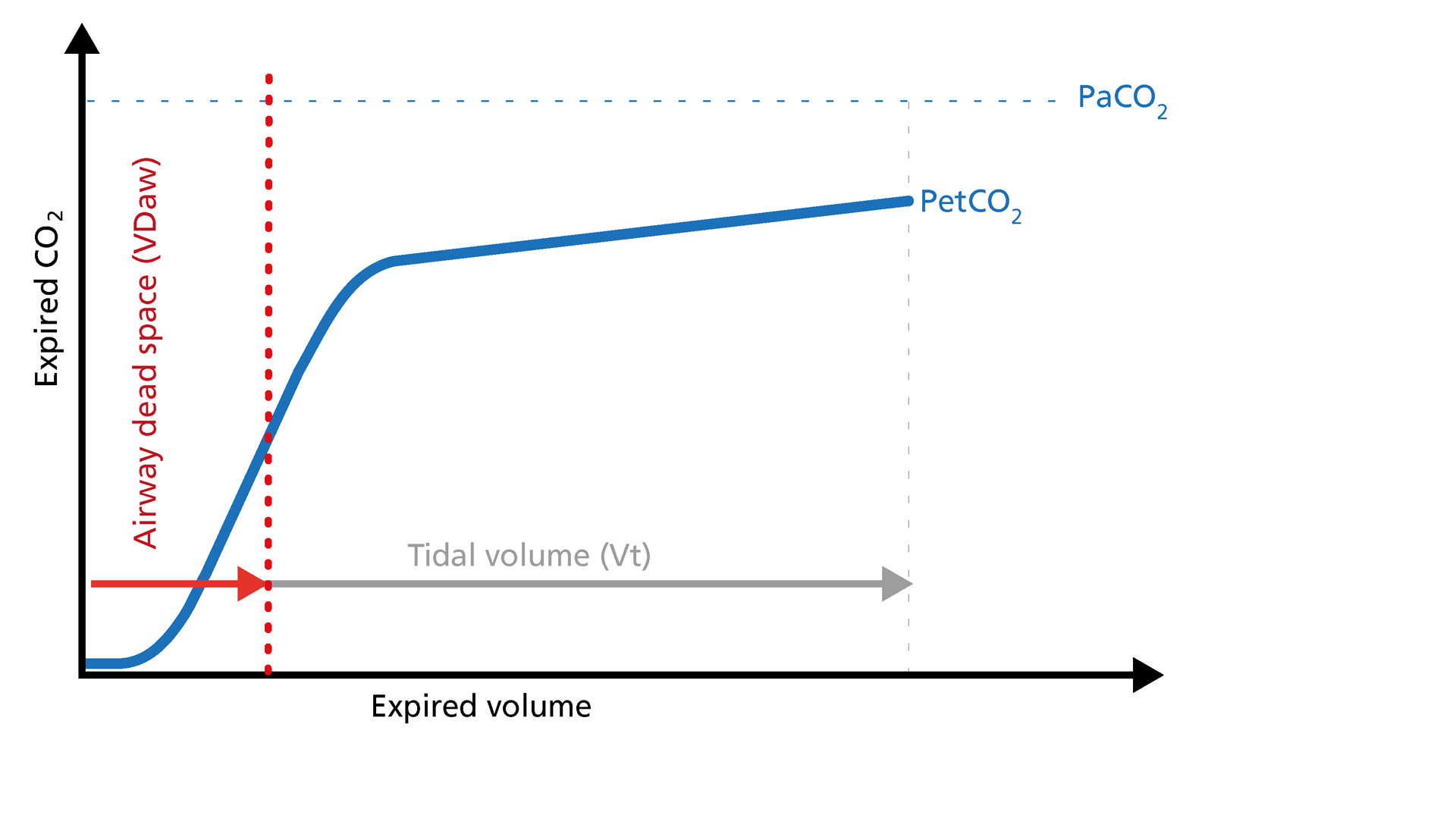
In the illustration (Figure 1) you can see:

  1. Slope of Phase III
  2. Slope of Phase II
  3. The intersection of lines 1 and 2 defines the limit between Phases II and III
  4. A perpendicular line is projected onto the X-axis and its position is adjusted until the areas p and q on both sides become equal
Illustration of the three areas of the volumetric capnogram
Figure 1: The three areas of the volumetric capnogram
Illustration of the three areas of the volumetric capnogram
Figure 1: The three areas of the volumetric capnogram

Area X – CO2 elimination (V‘CO2)

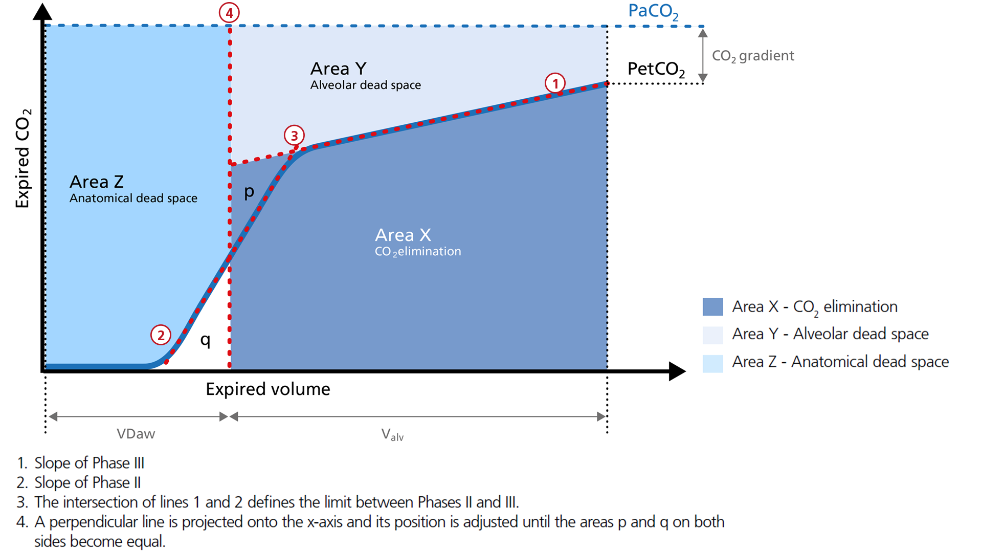
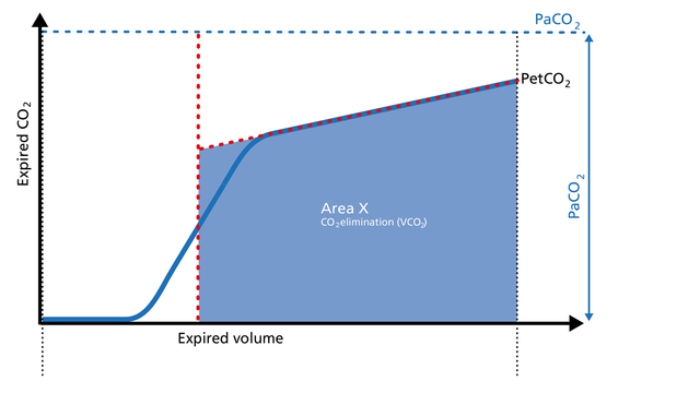
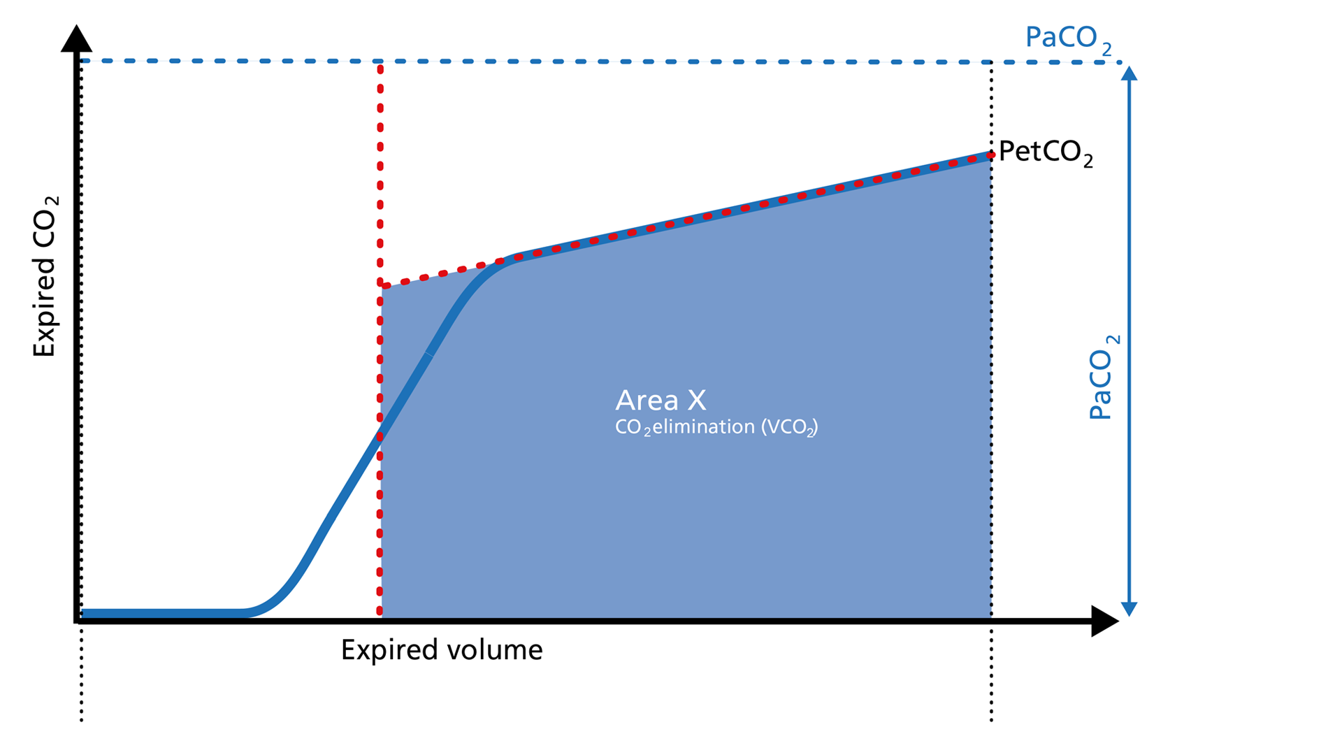
Area X represents the actual volume of CO2 exhaled in one breath (VeCO2). Adding up all of the single breaths in one minute gives you the total elimination of CO2 per minute (V‘CO2). If cardiac output, lung perfusion, and ventilation are stable, this is an assessment of the production of CO2 called V‘CO2. The V‘CO2 value displayed on the ventilator can be affected by any change in CO2 production, cardiac output, lung perfusion, and ventilation. It indicates instantly how the patient’s gas exchange responds to a change in ventilator settings. Monitoring trends allows for detection of sudden and rapid changes in V‘CO2 (Figure 2).

Good to know:

Decreasing V‘CO2: Hypothermia, deep sedation, hypothyroidism, paralysis, and brain death decrease CO2 production and induce a decrease in V‘CO2. Decreasing V‘CO2 can also be due to a decrease in cardiac output or blood loss, and may also suggest a change in blood flow to the lung areas. Pulmonary embolism, for example, exhibits V‘CO2 reduction and a slope reduction in Phase II.

Increase in V‘CO2: An increase in V'CO2 is usually due to bicarbonate infusion or an increase in CO2 production that can be caused by:

  • Fever
  • Sepsis
  • Seizures
  • Hyperthyroidism
  • Insulin therapy
Illustration of the Area X of the volumetric capnogram
Figure 2: Area X of the volumetric capnogram
Illustration of the Area X of the volumetric capnogram
Figure 2: Area X of the volumetric capnogram

Area Y - Alveolar dead space

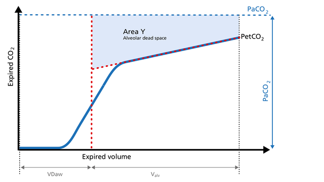
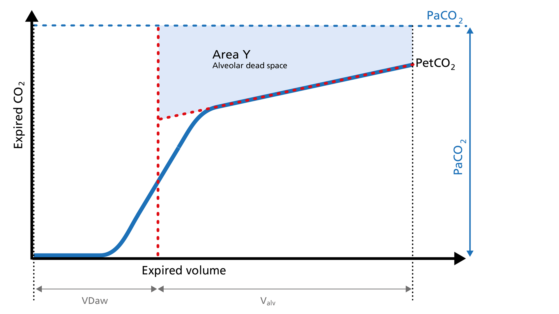
Area Y represents the amount of CO2 that is not eliminated due to alveolar dead space (Figure 3).

Good to know:

Increase: Alveolar dead space is increased in cases of lung emphysema, lung overdistension, pulmonary embolism, pulmonary hypertension, and cardiac output compromise.

Decrease: If the above mentioned conditions improve due to successful therapy, the alveolar
dead space decreases.

Illustration of the Area Y of the volumetric capnogram
Figure 3: Area Y of the volumetric capnogram
Illustration of the Area Y of the volumetric capnogram
Figure 3: Area Y of the volumetric capnogram

Area Z - Anatomical dead space

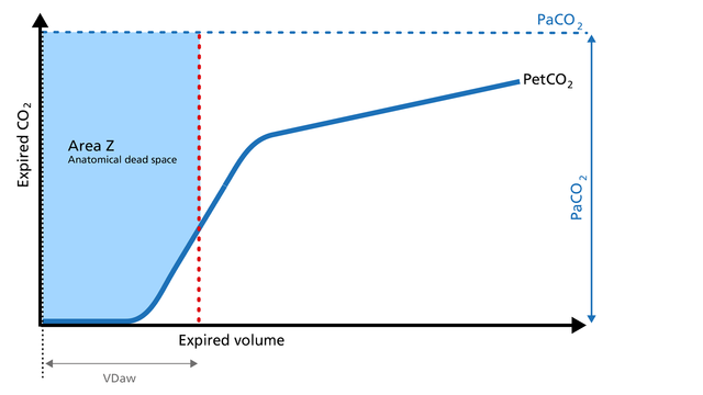
Anatomical dead space measurement using a volumetric capnogram gives an effective, in-vivo measure of volume lost in the conducting airway. This area represents a volume without CO2. It does not take part in the gas exchange and consists of the airway, endotracheal tube, and artificial accessories, such as a flextube positioned between the CO2 sensor and the patient (Figure 4).

Good to know:

Expansion of Area Z: An expansion of Area Z can indicate an increase in anatomical dead space ventilation (VDaw). Consider a reduction in your artificial dead space volume.

Diminution of Area Z: A diminution of Area Z is seen when the artificial dead space volume is decreased and when excessive PEEP is decreased.

Illustration of the Area Z of the volumetric capnogram
Figure 4: Area Z of the volumetric capnogram
Illustration of the Area Z of the volumetric capnogram
Figure 4: Area Z of the volumetric capnogram

Alveolar minute ventilation – V‘alv

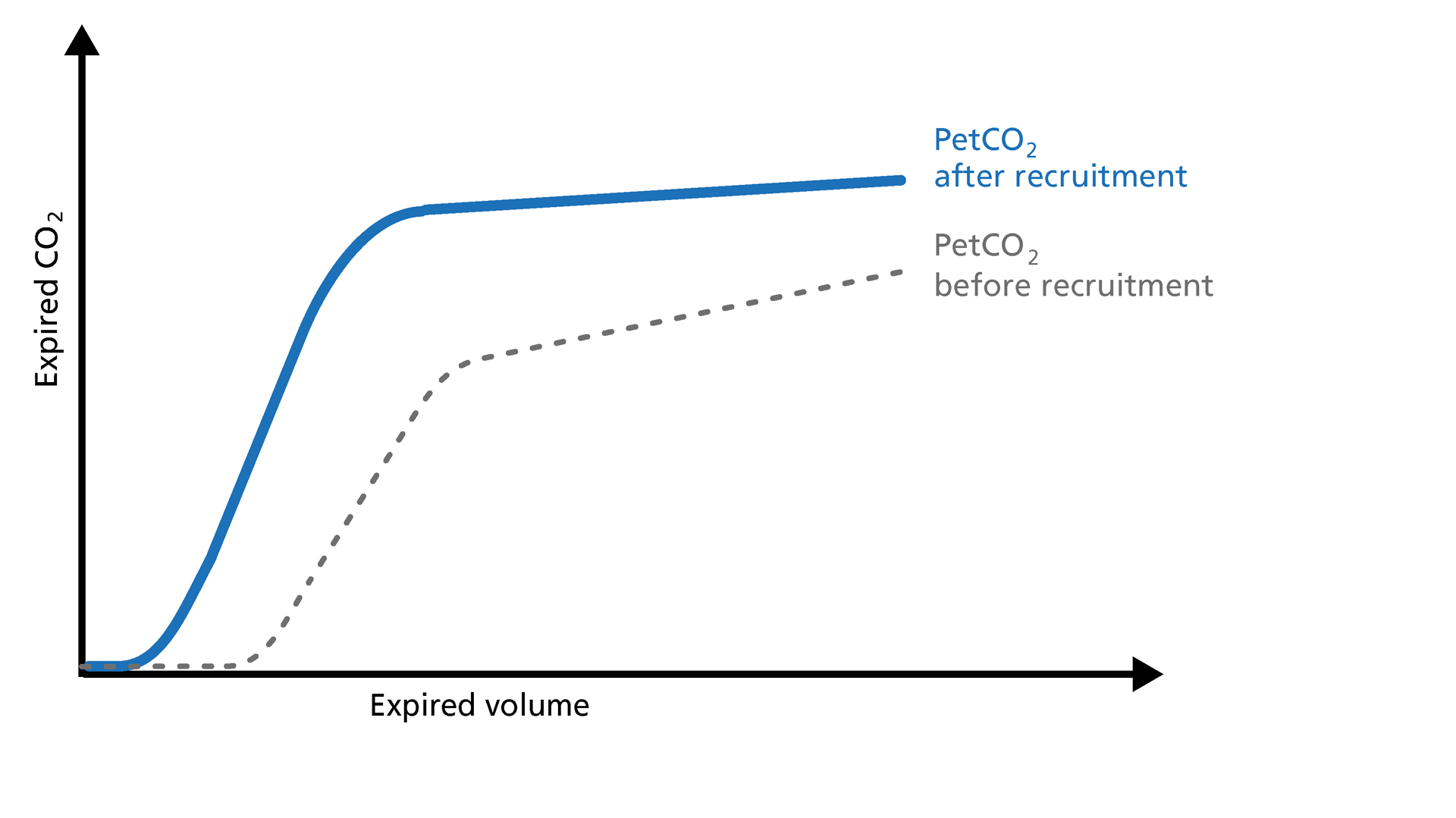
Phase III of the waveform represents the quantity of gas that comes from the alveoli and actively participates in gas exchange. V‘alv is calculated by subtracting the anatomical dead space (VDaw) from the tidal volume (VTE) multiplied by the respiratory rate from the minute volume (MinVol): V’alv =RR*Vtalv = RR*(VTE-VDaw) (Figure 5).

Good to know:

Increase: An increase in V‘alv is seen after an efficient recruitment maneuver and induces a transient increase in V‘CO2.
Decrease: A decrease in V‘alv can indicate that fewer alveoli are participating in the gas exchange, for example, due to pulmonary edema.

Illustration of PetCO2 before and after recruitment
Figure 5: PetCO2 before and after recruitment
Illustration of PetCO2 before and after recruitment
Figure 5: PetCO2 before and after recruitment

Dead space ventilation - VDaw/VTE ratio

The ratio of airway dead space (VDaw) to tidal volume (VTE) – the VDaw/VTE ratio – gives you an insight into the effectiveness of ventilation (Figure 6).

Good to know: A rising VDaw/VTE ratio can be a sign of ARDS.

  • In a normal lung, the VDaw/VTE ratio is between 25% and 30%.
  • In early ARDS, it is between 58% and up to 83%.
Illustration of the dead space ventilation on the volumetric capnogram
Figure 6: Dead space ventilation
Illustration of the dead space ventilation on the volumetric capnogram
Figure 6: Dead space ventilation

Volumetric capnography on Hamilton Medical ventilators

All Hamilton Medical ventilators offer volumetric capnography (All models except HAMILTON-MR1A​). It is available as an option on the HAMILTON‑C6, the HAMILTON‑G5, the HAMILTON‑C3, and the HAMILTON‑C1/T1, and as a standard feature on the HAMILTON‑S1.


Full citations below: (Anderson JT, Owings JT, Goodnight JE. Bedside noninvasive detection of acute pulmonary embolism in critically ill surgical patients. Arch Surg. 1999;134(8):869-875. doi:10.1001/archsurg.134.8.8691​), (Aström E, Niklason L, Drefeldt B, Bajc M, Jonson B. Partitioning of dead space--a method and reference values in the awake human. Eur Respir J. 2000;16(4):659-664. doi:10.1034/j.1399-3003.2000.16d16.x2​), (Blanch L, Romero PV, Lucangelo U. Volumetric capnography in the mechanically ventilated patient. Minerva Anestesiol. 2006;72(6):577-585. 3​), (Eriksson L, Wollmer P, Olsson CG, et al. Diagnosis of pulmonary embolism based upon alveolar dead space analysis. Chest. 1989;96(2):357-362. doi:10.1378/chest.96.2.3574​), (Fletcher R, Jonson B, Cumming G, Brew J. The concept of deadspace with special reference to the single breath test for carbon dioxide. Br J Anaesth. 1981;53(1):77-88. doi:10.1093/bja/53.1.775​), (Kallet RH, Daniel BM, Garcia O, Matthay MA. Accuracy of physiologic dead space measurements in patients with acute respiratory distress syndrome using volumetric capnography: comparison with the metabolic monitor method. Respir Care. 2005;50(4):462-467. 6​), (Kiiski R, Takala J. Hypermetabolism and efficiency of CO2 removal in acute respiratory failure. Chest. 1994;105(4):1198-1203. doi:10.1378/chest.105.4.11987​), (Kumar AY, Bhavani-Shankar K, Moseley HS, Delph Y. Inspiratory valve malfunction in a circle system: pitfalls in capnography. Can J Anaesth. 1992;39(9):997-999. doi:10.1007/BF030083538​) , (Nuckton TJ, Alonso JA, Kallet RH, et al. Pulmonary dead-space fraction as a risk factor for death in the acute respiratory distress syndrome. N Engl J Med. 2002;346(17):1281-1286. doi:10.1056/NEJMoa0128359​), (Eriksson L, Wollmer P, Olsson CG, et al. Diagnosis of pulmonary embolism based upon alveolar dead space analysis. Chest. 1989;96(2):357-362. doi:10.1378/chest.96.2.35710​), (Pyles ST, Berman LS, Modell JH. Expiratory valve dysfunction in a semiclosed circle anesthesia circuit--verification by analysis of carbon dioxide waveform. Anesth Analg. 1984;63(5):536-537. 11​), (RADFORD EP Jr. Ventilation standards for use in artificial respiration. J Appl Physiol. 1955;7(4):451-460. doi:10.1152/jappl.1955.7.4.45112​), (Rodger MA, Jones G, Rasuli P, et al. Steady-state end-tidal alveolar dead space fraction and D-dimer: bedside tests to exclude pulmonary embolism. Chest. 2001;120(1):115-119. doi:10.1378/chest.120.1.11513​) , (Yaron M, Padyk P, Hutsinpiller M, Cairns CB. Utility of the expiratory capnogram in the assessment of bronchospasm. Ann Emerg Med. 1996;28(4):403-407. doi:10.1016/s0196-0644(96)70005-714​), (Wolff G, Brunner JX, Grädel E. Gas exchange during mechanical ventilation and spontaneous breathing. Intermittent mandatory ventilation after open heart surgery. Chest. 1986;90(1):11-17. doi:10.1378/chest.90.1.1115​), (Wolff G, X. B. J. , Weibel W., Bowes C.L. , Muchenberger R., Bertschmann W. (1989). Anatomical and series dead space volume: concept and measurement in clinical practice. Applied cardiopulmonary pathophysiology, 2, 299-307.16)

Volumentric Capnography e-book

Free e-book

Good to know! All about volumetric capnography

Learn how to interpret a volumetric capnogram and get an overview of the benefits and clinical applications of volumetric capnography. Includes a self-test!
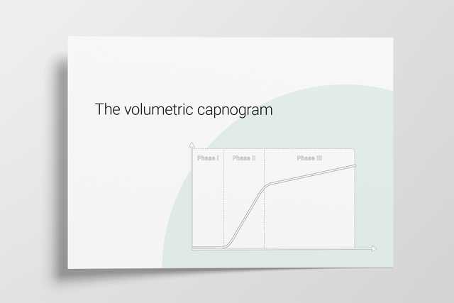
Footnotes

  • A. All models except HAMILTON-MR1

References

  1. 1. Anderson JT, Owings JT, Goodnight JE. Bedside noninvasive detection of acute pulmonary embolism in critically ill surgical patients. Arch Surg. 1999;134(8):869-875. doi:10.1001/archsurg.134.8.869
  2. 2. Aström E, Niklason L, Drefeldt B, Bajc M, Jonson B. Partitioning of dead space--a method and reference values in the awake human. Eur Respir J. 2000;16(4):659-664. doi:10.1034/j.1399-3003.2000.16d16.x
  3. 3. Blanch L, Romero PV, Lucangelo U. Volumetric capnography in the mechanically ventilated patient. Minerva Anestesiol. 2006;72(6):577-585.
  4. 4. Eriksson L, Wollmer P, Olsson CG, et al. Diagnosis of pulmonary embolism based upon alveolar dead space analysis. Chest. 1989;96(2):357-362. doi:10.1378/chest.96.2.357
  5. 5. Fletcher R, Jonson B, Cumming G, Brew J. The concept of deadspace with special reference to the single breath test for carbon dioxide. Br J Anaesth. 1981;53(1):77-88. doi:10.1093/bja/53.1.77
  6. 6. Kallet RH, Daniel BM, Garcia O, Matthay MA. Accuracy of physiologic dead space measurements in patients with acute respiratory distress syndrome using volumetric capnography: comparison with the metabolic monitor method. Respir Care. 2005;50(4):462-467.
  7. 7. Kiiski R, Takala J. Hypermetabolism and efficiency of CO2 removal in acute respiratory failure. Chest. 1994;105(4):1198-1203. doi:10.1378/chest.105.4.1198
  8. 8. Kumar AY, Bhavani-Shankar K, Moseley HS, Delph Y. Inspiratory valve malfunction in a circle system: pitfalls in capnography. Can J Anaesth. 1992;39(9):997-999. doi:10.1007/BF03008353
  9. 9. Nuckton TJ, Alonso JA, Kallet RH, et al. Pulmonary dead-space fraction as a risk factor for death in the acute respiratory distress syndrome. N Engl J Med. 2002;346(17):1281-1286. doi:10.1056/NEJMoa012835
  10. 10. Olsson K, Jonson B, Olsson CG, Wollmer P. Diagnosis of pulmonary embolism by measurement of alveolar dead space. J Intern Med. 1998;244(3):199-207. doi:10.1046/j.1365-2796.1998.00356.x
  11. 11. Pyles ST, Berman LS, Modell JH. Expiratory valve dysfunction in a semiclosed circle anesthesia circuit--verification by analysis of carbon dioxide waveform. Anesth Analg. 1984;63(5):536-537.
  12. 12. RADFORD EP Jr. Ventilation standards for use in artificial respiration. J Appl Physiol. 1955;7(4):451-460. doi:10.1152/jappl.1955.7.4.451
  13. 13. Rodger MA, Jones G, Rasuli P, et al. Steady-state end-tidal alveolar dead space fraction and D-dimer: bedside tests to exclude pulmonary embolism. Chest. 2001;120(1):115-119. doi:10.1378/chest.120.1.115
  14. 14. Yaron M, Padyk P, Hutsinpiller M, Cairns CB. Utility of the expiratory capnogram in the assessment of bronchospasm. Ann Emerg Med. 1996;28(4):403-407. doi:10.1016/s0196-0644(96)70005-7
  15. 15. Wolff G, Brunner JX, Grädel E. Gas exchange during mechanical ventilation and spontaneous breathing. Intermittent mandatory ventilation after open heart surgery. Chest. 1986;90(1):11-17. doi:10.1378/chest.90.1.11
  16. 16). Wolff G, X. B. J. , Weibel W., Bowes C.L. , Muchenberger R., Bertschmann W. (1989). Anatomical and series dead space volume: concept and measurement in clinical practice. Applied cardiopulmonary pathophysiology, 2, 299-307.

Bedside noninvasive detection of acute pulmonary embolism in critically ill surgical patients.

Anderson JT, Owings JT, Goodnight JE. Bedside noninvasive detection of acute pulmonary embolism in critically ill surgical patients. Arch Surg. 1999;134(8):869-875. doi:10.1001/archsurg.134.8.869


HYPOTHESIS

We hypothesized that late pulmonary dead space fraction (Fd(late)) would be a useful tool to screen for pulmonary embolism (PE) in a group of surgical patients, including patients who required mechanical ventilation and patients with adult respiratory distress syndrome.

DESIGN

We prospectively calculated Fd(late) in patients with suspected PE who underwent pulmonary angiography.

SETTING

University-based, level I trauma center.

MAIN OUTCOME MEASURE

Ability of Fd(late) to identify patients with PE.

RESULTS

Twelve patients had 14 angiograms for suspected PE. The Fd(late) was 0.12 or above in all 5 patients who had PE; 4 required mechanical ventilation. The Fd(late) values were below 0.12 in 8 of 9 patients without PE. Four patients had adult respiratory distress syndrome. The Fd(late) had 100% sensitivity and 89% specificity for the detection of PE.

CONCLUSIONS

The Fd(late) is a valuable tool for bedside screening of PE in surgical patients. We were able to accurately detect all PEs.

Partitioning of dead space--a method and reference values in the awake human.

Aström E, Niklason L, Drefeldt B, Bajc M, Jonson B. Partitioning of dead space--a method and reference values in the awake human. Eur Respir J. 2000;16(4):659-664. doi:10.1034/j.1399-3003.2000.16d16.x

Although dead space is often increased in disease, it is not frequently measured in the clinic. This may reflect that an adequate method as well as reference values are missing. Healthy males and females, n=38, age 20-61 yrs, were connected to a pneumotachograph and a fast CO2 analyser after radial artery catheterization. The physiological dead space was partitioned into airway and alveolar dead space using a delineation principle denoted the pre-interface expirate. Physiological dead space was 201+/-41 mL in males and 150+/-34 mL in females. Dead space values were depending upon parameters reflecting lung size (predicted total lung capacity), breathing pattern and age. After multiple correlation the variation decreased and differences between males and females disappeared. The residual SD was then for physiological dead space 18.9 mL. The clinical use of the new method for determination of dead space can be based upon reference values, with a more narrow range than previous data.

Volumetric capnography in the mechanically ventilated patient.

Blanch L, Romero PV, Lucangelo U. Volumetric capnography in the mechanically ventilated patient. Minerva Anestesiol. 2006;72(6):577-585.

Expiratory capnogram provides qualitative information on the waveform patterns associated with mechanical ventilation and quantitative estimation of expired CO2. Volumetric capnography simultaneously measures expired CO2 and tidal volume and allows identification of CO2 from 3 sequential lung compartments: apparatus and anatomic dead space, from progressive emptying of alveoli and alveolar gas. Lung heterogeneity creates regional differences in CO2 concentration and sequential emptying contributes to the rise of the alveolar plateau and to the steeper the expired CO2 slope. The concept of dead space accounts for those lung areas that are ventilated but not perfused. In patients with sudden pulmonary vascular occlusion due to pulmonary embolism, the resultant high V/Q mismatch produces an increase in alveolar dead space. Calculations derived from volumetric capnography are useful to suspect pulmonary embolism at the bedside. Alveolar dead space is large in acute lung injury and when the effect of positive end-expiratory pressure (PEEP) is to recruit collapsed lung units resulting in an improvement of oxygenation, alveolar dead space may decrease, whereas PEEP-induced overdistension tends to increase alveolar dead space. Finally, measurement of physiologic dead space and alveolar ejection volume at admission or the trend during the first 48 hours of mechanical ventilation might provide useful information on outcome of critically ill patients with acute lung injury or acute respiratory distress syndrome.

Diagnosis of pulmonary embolism based upon alveolar dead space analysis.

Eriksson L, Wollmer P, Olsson CG, et al. Diagnosis of pulmonary embolism based upon alveolar dead space analysis. Chest. 1989;96(2):357-362. doi:10.1378/chest.96.2.357

Pulmonary embolism (PE) leads to an abnormal alveolar deadspace that is expired in synchrony with gas from normally perfused alveoli. This feature of PE separates it from pulmonary diseases affecting the airways, which are characterized by nonsynchronous emptying of compartments with an uneven ventilation/perfusion relationship. An analysis of the single breath test (SBT) for CO2, SBT-CO2, focusing on the late tidal expirate, was made in order to evaluate the feasibility to use the SBT-CO2 for the diagnosis of PE. The test was evaluated in 38 patients with suspected PE where pulmonary angiography showed that nine had PE and 29 did not. It was also tested in a reference population consisting of patients with normal lung function, obstructive lung disease and interstitial lung disease. Previously suggested gas exchange measurements for the diagnosis of PE, ie, the physiologic deadspace fraction, VDphys/VT, and the arterial-to-end-tidal CO2 gradient, P(a-E')CO2, were also evaluated in the groups. SBT-CO2 achieved a nearly complete separation between the patients with PE and those without. The other measurements, however, showed a substantial overlap between patients with PE and those with obstructive or interstitial lung disease. The SBT-CO2 is simple and potentially widely available and warrants further study as a routine technique for the diagnosis of PE.

The concept of deadspace with special reference to the single breath test for carbon dioxide.

Fletcher R, Jonson B, Cumming G, Brew J. The concept of deadspace with special reference to the single breath test for carbon dioxide. Br J Anaesth. 1981;53(1):77-88. doi:10.1093/bja/53.1.77

We present a review and a theoretical analysis of factors determining airway deadspace (VDaw) and alveolar deadspace (VDalv), the two constituents of physiological deadspace (VDphys). VDaw if the volume of gas between the lips and the alveolar/fresh gas interface, the location of which is determined by inspiratory flow pattern and airway geometry. VDalv can be caused by incomplete alveolar gas mixing and associated V/Q mismatching within the terminal respiratory units, temporal V/Q mismatching within units, spatial V/Q mismatching between units, and venous admixture. Most causes of VDphys are influenced by inspiratory flow pattern and the time available for gas diffusion and distribution. Analysis can be made from the single breath test for carbon dioxide (SBT--CO2) which is the plot of fraction of carbon dioxide in expired gas against expired volume. The common causes of VDalv are associated with a sloping SBT-CO2 phase III. Combination of SBT-CO2 with PaCO2 yields VDphys and VDalv. A sloping phase III with a negative arterial-end-tidal PCO2 gradient implies compensation by perfusion for early emptying, overventilated alveoli.

Accuracy of physiologic dead space measurements in patients with acute respiratory distress syndrome using volumetric capnography: comparison with the metabolic monitor method.

Kallet RH, Daniel BM, Garcia O, Matthay MA. Accuracy of physiologic dead space measurements in patients with acute respiratory distress syndrome using volumetric capnography: comparison with the metabolic monitor method. Respir Care. 2005;50(4):462-467.


BACKGROUND

Volumetric capnography is an alternative method of measuring expired carbon dioxide partial pressure (P(eCO2)) and physiologic dead-space-to-tidal-volume ratio (V(D)/V(T)) during mechanical ventilation. In this method, P(eCO2) is measured at the Y-adapter of the ventilator circuit, thus eliminating the effects of compression volume contamination and the need to apply a correction factor. We investigated the accuracy of volumetric capnography in measuring V(D)/V(T), compared to both uncorrected and corrected measurements, using a metabolic monitor in patients with acute respiratory distress syndrome (ARDS).

METHODS

There were 90 measurements of V(D)/V(T) made in 23 patients with ARDS. The P(eCO2) was measured during a 5-min expired-gas collection period with a Delta-trac metabolic monitor, and was corrected for compression volume contamination using a standard formula. Simultaneous measurements of P(eCO2) and V(D)/V(T) were obtained using volumetric capnography.

RESULTS

V(D)/V(T) measured by volumetric capnography was strongly correlated with both the uncorrected (r2 = 0.93, p < 0.0001) and corrected (r2 = 0.89, p < 0.0001) measurements of V(D)/V(T) made using the metabolic monitor technique. Measurements of V(D)/V(T) made with volumetric capnography had a bias of 0.02 and a precision of 0.05 when compared to the V(D)/V(T) corrected for estimated compression volume contamination.

CONCLUSION

Volumetric capnography measurements of V(D)/V(T) in mechanically-ventilated patients with ARDS are as accurate as those obtained by metabolic monitor technique. .

Hypermetabolism and efficiency of CO2 removal in acute respiratory failure.

Kiiski R, Takala J. Hypermetabolism and efficiency of CO2 removal in acute respiratory failure. Chest. 1994;105(4):1198-1203. doi:10.1378/chest.105.4.1198


OBJECTIVE

To assess the effect of hypermetabolism, dead-space ventilation, and parenteral nutrition on the minute ventilation requirement in mechanically ventilated patients.

DESIGN

A retrospective analysis of data collected in study protocols unrelated to the present study.

SETTING

A medical-surgical intensive care unit in a tertiary care center.

PATIENTS

One hundred eleven mechanically ventilated patients were studied during volume-controlled ventilation.

MEASUREMENTS

Gas exchange measurement by indirect calorimetry and arterial blood gas analysis.

METHODS

Minute ventilation (VE), carbon dioxide production (VCO2), and respiratory exchange ratio (RER) were measured with indirect calorimetry. Arterial CO2 tension was sampled at the end of the measurement, and alveolar ventilation (VA), deadspace to tidal volume ratio (VD/VT) and predicted resting VCO2 were calculated. The VE demand at a standard PaCO2 was calculated and the contribution of the observed hypermetabolism and increased VD/VT was identified. In a subgroup of patients, the effect of initiating parenteral nutrition on the VE demand was assessed. There were four study groups: multiple trauma, sepsis, ARDS, and postoperative open-heart surgery patients.

MAIN RESULTS

A combination of hypermetabolism and increased dead-space was observed in 67 of the 111 patients. Increased VCO2 accounted for 69 percent of the excess VE demand in trauma, 67 percent in sepsis, 58 percent in postoperative patients, and 56 percent in ARDS. Parenteral nutrition with a caloric intake matching measured resting energy expenditure (REE) did not increase VCO2 or the demand for VE.

CONCLUSIONS

Increased VCO2 is the main cause of increased VE demand in the majority of mechanically ventilated ICU patients. Parenteral nutrition at energy intakes close to actual REE does not increase the ventilatory demand.

Inspiratory valve malfunction in a circle system: pitfalls in capnography.

Kumar AY, Bhavani-Shankar K, Moseley HS, Delph Y. Inspiratory valve malfunction in a circle system: pitfalls in capnography. Can J Anaesth. 1992;39(9):997-999. doi:10.1007/BF03008353

Capnography is a useful technique in monitoring the integrity of anaesthetic equipment such as the malfunctioning of unidirectional valves in circle system. However, the lack of a precise mechanism in existing capnographs to identify the start of inspiration and the beginning of expiration in the capnograms, makes the analysis of the carbon dioxide waveforms during inspiration difficult and thus results in inaccurate assessment of rebreathing. We report a case where, during the malfunction of the inspiratory unidirectional valve in the circle system, the capnograph failed to detect the presence of substantial rebreathing. Critical analysis of the capnogram recorded during the malfunction revealed that there was substantial rebreathing which was underestimated by the capnograph as it reports only the lowest CO2 concentration rebreathed during inspiration in such abnormal situations.

Pulmonary dead-space fraction as a risk factor for death in the acute respiratory distress syndrome.

Nuckton TJ, Alonso JA, Kallet RH, et al. Pulmonary dead-space fraction as a risk factor for death in the acute respiratory distress syndrome. N Engl J Med. 2002;346(17):1281-1286. doi:10.1056/NEJMoa012835


BACKGROUND

No single pulmonary-specific variable, including the severity of hypoxemia, has been found to predict the risk of death independently when measured early in the course of the acute respiratory distress syndrome. Because an increase in the pulmonary dead-space fraction has been described in observational studies of the syndrome, we systematically measured the dead-space fraction early in the course of the illness and evaluated its potential association with the risk of death.

METHODS

The dead-space fraction was prospectively measured in 179 intubated patients, a mean (+/-SD) of 10.9+/-7.4 hours after the acute respiratory distress syndrome had developed. Additional clinical and physiological variables were analyzed with the use of multiple logistic regression. The study outcome was mortality before hospital discharge.

RESULTS

The mean dead-space fraction was markedly elevated (0.58+/-0.09) early in the course of the acute respiratory distress syndrome and was higher among patients who died than among those who survived (0.63+/-0.10 vs. 0.54+/-0.09, P<0.001). The dead-space fraction was an independent risk factor for death: for every 0.05 increase, the odds of death increased by 45 percent (odds ratio, 1.45; 95 percent confidence interval, 1.15 to 1.83; P=0.002). The only other independent predictors of an increased risk of death were the Simplified Acute Physiology Score II, an indicator of the severity of illness (odds ratio, 1.06; 95 percent confidence interval, 1.03 to 1.08; P<0.001) and quasistatic respiratory compliance (odds ratio, 1.06; 95 percent confidence interval, 1.01 to 1.10; P=0.01).

CONCLUSIONS

Increased dead-space fraction is a feature of the early phase of the acute respiratory distress syndrome. Elevated values are associated with an increased risk of death.

Diagnosis of pulmonary embolism by measurement of alveolar dead space.

Olsson K, Jonson B, Olsson CG, Wollmer P. Diagnosis of pulmonary embolism by measurement of alveolar dead space. J Intern Med. 1998;244(3):199-207. doi:10.1046/j.1365-2796.1998.00356.x


OBJECTIVE

Pulmonary embolism (PE) gives rise to alveolar dead space, which can be measured with a single breath test for CO2 (SBT-CO2). The characteristics of the SBT-CO2 are different in PE and other common conditions giving rise to alveolar dead space, notably airways disease. An analysis of alveolar dead space focusing on the late part of the breath (fDlate) has been suggested as a method for diagnosis of PE. Our aim was to evaluate this technique by comparison with lung scintigraphy.

METHODS

We randomly selected patients with clinical suspicion of PE. SBT-CO2 and lung scintigraphy were performed on the same day. The scintigraphies were reviewed and classified as high, intermediate and low probability of PE.

RESULTS

Out of 223 patients able to be evaluated, there were 20 of the high, 29 of the intermediate and 174 of the low probability category. There were large differences between the means of fDlate in the high and the intermediate and in the high and the low categories. We obtained a sensitivity of 85% and a specificity of 93% for diagnosis of PE, based on high and low probability categories. If a patient with previous PE, but no scintigraphic evidence of current PE, is excluded the sensitivity increases to 90%.

CONCLUSIONS

This study provides further support for the measurement of fDlate by the SBT-CO2 as a diagnostic test in patients with suspicion of PE. The test should be especially useful in small hospitals without access to pulmonary scintigraphy or pulmonary angiography.

Expiratory valve dysfunction in a semiclosed circle anesthesia circuit--verification by analysis of carbon dioxide waveform.

Pyles ST, Berman LS, Modell JH. Expiratory valve dysfunction in a semiclosed circle anesthesia circuit--verification by analysis of carbon dioxide waveform. Anesth Analg. 1984;63(5):536-537.

Ventilation standards for use in artificial respiration.

RADFORD EP Jr. Ventilation standards for use in artificial respiration. J Appl Physiol. 1955;7(4):451-460. doi:10.1152/jappl.1955.7.4.451

Steady-state end-tidal alveolar dead space fraction and D-dimer: bedside tests to exclude pulmonary embolism.

Rodger MA, Jones G, Rasuli P, et al. Steady-state end-tidal alveolar dead space fraction and D-dimer: bedside tests to exclude pulmonary embolism. Chest. 2001;120(1):115-119. doi:10.1378/chest.120.1.115


STUDY OBJECTIVE

Less than 35% of patients suspected of having pulmonary embolism (PE) actually have PE. Safe bedside methods to exclude PE could save health-care resources and improve access to diagnostic testing for suspected PE. In patients with suspected PE, we sought to determine the sensitivity, specificity, and negative predictive value of (1) a steady-state end-tidal alveolar dead space fraction (AVDSf) of < 0.15, (2) a negative D-dimer result, and (3) the combination of a steady-state end-tidal AVDSf of < 0.15 and a negative D-dimer result.

STUDY DESIGN

Prospective cohort study.

SETTING

Tertiary-care center in Ottawa, Ontario, Canada.

PATIENTS

Consecutive inpatients, outpatients, and emergency department patients with suspected PE referred to the Departments of Nuclear Medicine or Radiology for investigation of suspected PE.

INTERVENTIONS AND MEASUREMENTS

All study patients had D-Dimer and alveolar dead space measurements prior to determining outcome (PE or no PE) with ventilation/perfusion scans and/or noninvasive leg vein imaging and/or pulmonary angiography.

RESULTS

Two hundred forty-six eligible and consenting patients underwent diagnostic imaging that excluded PE in 163 patients, diagnosed PE in 49 patients, and was indeterminant in 34 patients. A negative D-dimer result excluded PE with a sensitivity of 83.0% (95% confidence interval [CI], 69.2 to 92.4%), a negative predictive value of 91.2% (95% CI, 83.4 to 96.1%), and a specificity of 57.6%. A steady-state end-tidal AVDSf of < 0.15 excluded PE with a sensitivity of 79.5% (95% CI, 63.5 to 90.7%), a negative predictive value of 90.7% (95% CI, 82.5 to 95.9%), and a specificity of 70.3%. The combination of a negative D-dimer result and a steady-state end-tidal AVDSf of < 0.15 excluded PE with a sensitivity of 97.8% (95% CI, 88.5 to 99.9%), a negative predictive value of 98.0% (95% CI, 89.4 to 99.9%), and a specificity of 38.0%.

CONCLUSION

This simple combination of bedside tests may safely rule out PE without further diagnostic testing in large numbers of patients with suspected PE.

Utility of the expiratory capnogram in the assessment of bronchospasm.

Yaron M, Padyk P, Hutsinpiller M, Cairns CB. Utility of the expiratory capnogram in the assessment of bronchospasm. Ann Emerg Med. 1996;28(4):403-407. doi:10.1016/s0196-0644(96)70005-7


STUDY OBJECTIVE

To determine whether the plateau phase of the expiratory capnogram (dco2/dt) can detect bronchospasm in adult asthma patients in the emergency department and to assess the correlation between dco2/dt and the peak expiratory flow rate (PEFR) in spontaneously breathing patients with asthma and in normal, healthy volunteers.

METHODS

We carried out a prospective, blinded study in a university hospital ED. Twenty adults (12 women) with acute asthma and 28 normal adult volunteers (15 women) breathed through the sampling probe of an end-tidal CO2 monitor, and the expired CO2 waveform was recorded. The dco2/dt of the plateau (alveolar) phase for five consecutive regular expirations was measured and a mean value calculated for each patient. The best of three PEFRs was determined. The PEFR and dco2/dt were also recorded after treatment of the asthmatic patients with inhaled beta-agonists.

RESULTS

The mean +/- SD PEFR of the asthmatic subjects was 274 +/- 96 L/minute (57% of the predicted value), whereas that of the normal volunteers was 527 +/- 96 L/minute (103% of the predicted value) (P < .001). The mean dco2/dt of the asthmatic subjects (.26 +/- .06) was significantly steeper than that of the normal volunteers (.13 +/- .06) (P < .001). The dco2/dt was correlated with PEFR (r = .84, P < .001). In 18 asthmatic subjects the pretreatment and posttreatment percent predicted PEFRS were 58% +/- 17% and 74% +/- 17%, respectively (P < .001), whereas the dco2/dt values were .27 +/- .05 and .19 +/- .07, respectively (P < .005).

CONCLUSION

The dco2/dt is an effort-independent, rapid noninvasive measure that indicates significant bronchospasm in ED adult patients with asthma. The dco2/dt value is correlated with PEFR, an effort-dependent measure of airway obstruction. The change in dco2/dt with inhaled beta-agonists may be useful in monitoring the therapy of acute asthma.

Gas exchange during mechanical ventilation and spontaneous breathing. Intermittent mandatory ventilation after open heart surgery.

Wolff G, Brunner JX, Grädel E. Gas exchange during mechanical ventilation and spontaneous breathing. Intermittent mandatory ventilation after open heart surgery. Chest. 1986;90(1):11-17. doi:10.1378/chest.90.1.11

Pulmonary gas exchange rates in eight patients after open heart surgery were studied during weaning from the ventilator. We investigated continuous positive pressure ventilation (CPPV), intermittent mandatory ventilation (IMV) and spontaneous breathing with continuous positive airway pressure (CPAP). During each mode of ventilation we measured: CO2 production (VCO2), O2 consumption (VO2), cardiac output (CO), PaO2, Qs/QT and functional residual capacity (FRC). In addition, we analyzed in each single breath: tidal volume (VT), series dead space volume (Vds), alveolar ventilation, alveolar efficiency for CO2 elimination (alv eff CO2) and end-tidal CO2 concentration (FCO2et). We compared the results of CPPV, IMV and CPAP and the mandatory breaths (MB) with the spontaneous breaths (SB) measured during IMV. CO was low during CPPV, when the patient still deeply sedated; it was increased in IMV and remained constant in the following CPAP period. VCO2 and VO2 did not differ significantly when switching from IMV to CPAP; therefore, work due to breathing seemed not to be reduced by the mandatory breath during IMV. Oxygenation (PaO2, Qs/QT) did not change significantly when switching from one mode to the other. FRC was constant when changing from CPPV to IMV, did not alter within the IMV-cycle and was reduced significantly when switching from IMV to CPAP. Dead space ventilation was reduced in SB (compared to MB). The latter result is discussed on the basis of two mechanisms: Vds was reduced and alv eff CO2 was increased. We conclude that compared to CPPV, IMV decreases mean alveolar pressure and reduces dead space ventilation at constant FRC and with constant oxygenation. This may explain why, in the weaning process, IMV makes it possible to start spontaneous breathing very early.

Anatomical and series dead space volume: concept and measurement in clinical practice

Wolff G, X. B. J. , Weibel W., Bowes C.L. , Muchenberger R., Bertschmann W. (1989). Anatomical and series dead space volume: concept and measurement in clinical practice. Applied cardiopulmonary pathophysiology, 2, 299-307.

Related articles. Get a deeper look