Back

Using waveforms to identify asynchronies - Step 2

Article

Author: Caroline Brown, Branka Cupic

Date of first publication: 30.09.2022

In the previous issue, our Bedside tip covered the starting point for identifying asynchronies using ventilator waveforms.

Using waveforms to identify asynchronies - Step 2

The first step is to know what a normal breath looks like and how to recognize good synchrony between the patient and ventilator. We looked at the concept of exponential decay, and how to identify the start of an inspiratory effort and the well-synchronized end of inspiration.

The next step is how to identify the most common minor and major asynchronies.

Before looking more closely at those, we will recap the principles that formed the basis for a systematic method of waveform analysis applied by Mojoli et al. (Mojoli F, Pozzi M, Orlando A, et al. Timing of inspiratory muscle activity detected from airway pressure and flow during pressure support ventilation: the waveform method. Crit Care. 2022;26(1):32. Published 2022 Jan 30. doi:10.1186/s13054-022-03895-41​):

  • In a patient with a normal breathing pattern, inspiration is active and expiration is passive
  • Exponential decay of flow indicates a passive condition (for both inspiratory and expiratory flow)
  • In the case of synchronous pressure-support ventilation, passive conditions should only be observed during the ventilator’s expiratory phase
  • Passive conditions during the ventilator’s inspiratory phase indicate auto-triggering or delayed cycling
  • Deviations from passive conditions during the ventilator’s expiratory phase indicate early cycling, ineffective inspiratory efforts, or expiratory muscle activation

Minor asynchrony - Early cycling

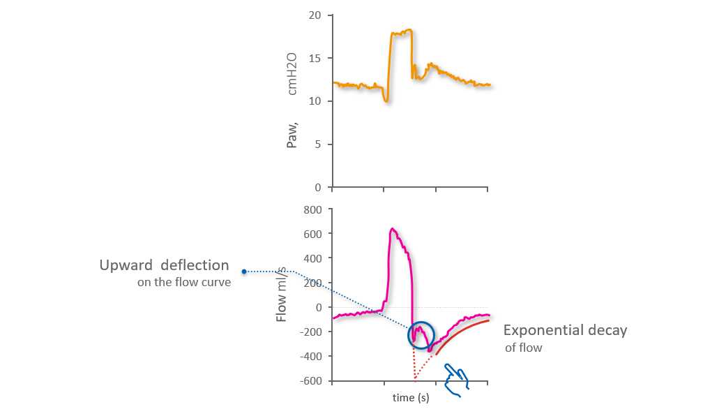
If patient and ventilator are synchronized and cycling is optimal, the Flow waveform will show the peak expiratory flow at the beginning of expiration, followed by exponential decay. Early cycling, whereby expiration starts before the inspiratory effort has ended, is denoted by a distortion of the initial expiratory flow: the normal peak flow is replaced by an upwards deflection, while the normal exponential decay starts later (see Figure 1 below).

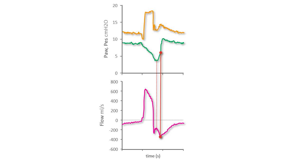
Using the esophageal pressure (Pes) waveform as a reference, we can see that the nadir of Pes (i.e., the point of maximum inspiratory effort) takes place during expiration and corresponds with the upwards deflection of the expiratory flow. The exponential flow decay starts later, namely at the end of the inspiratory effort as shown on the Pes waveform (see Figure 2 below). From our previous Bedside tip, we know that the fast increase in Pes after its nadir indicates relaxation of the inspiratory muscles, and the midpoint of this fast increase can be used as the reference for the end of inspiration.

Waveform showing upwards deflection
Figure 1 (Image modified from Mojoli et al. Critical Care (2022) 26:32)
Waveform showing upwards deflection
Figure 1 (Image modified from Mojoli et al. Critical Care (2022) 26:32)
Waveform showing late start of exponential decay
Figure 2 (Image modified from Mojoli et al. Critical Care (2022) 26:32)
Waveform showing late start of exponential decay
Figure 2 (Image modified from Mojoli et al. Critical Care (2022) 26:32)

Minor asynchrony - Delayed cycling

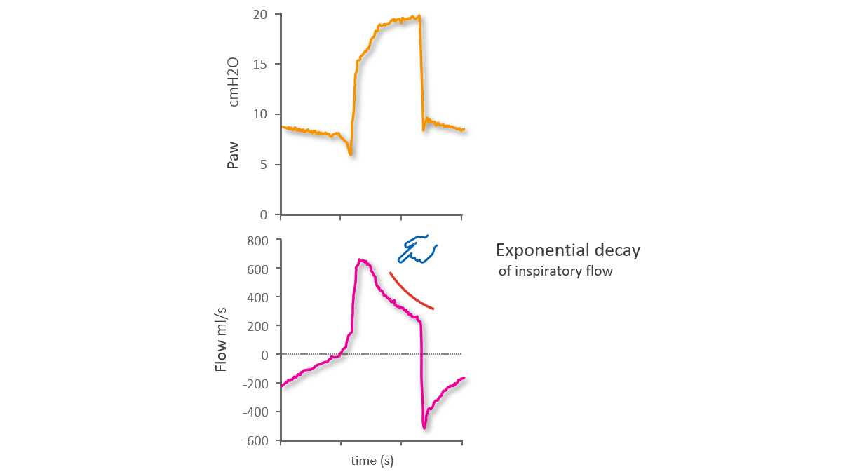
In the case of delayed cycling to expiration, there are two phases of inspiration. The first phase  is caused by the normal synchronized  pull of the inspiratory muscles and push from the ventilator, but inspiration then continues solely due to the push from the ventilator. This results in passive inflation. You can recognize this on the Flow waveform from the prolonged exponential decay of inspiratory flow occurring after the first phase with upwards convexity (see Figure 3 below).

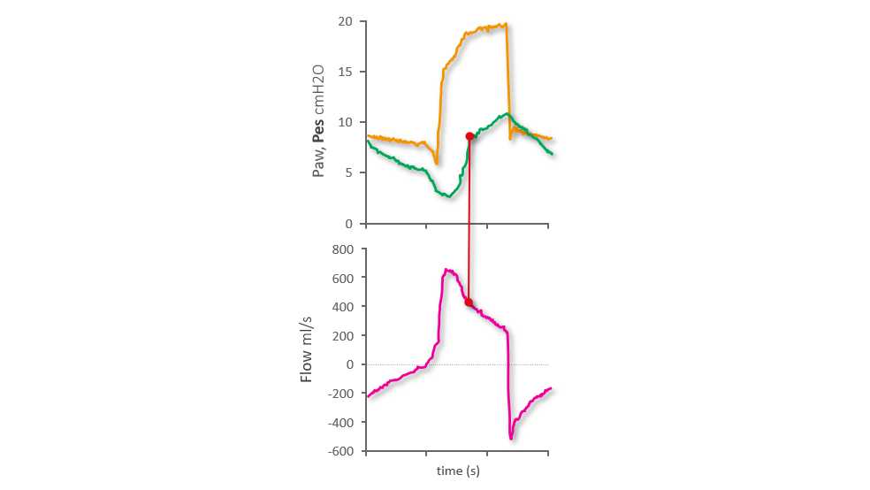
On the Pes waveform we can see a point on the Pes rise after its nadir that marks a clear change of slope. This point corresponds with the transition from synchronized inspiration to the subsequent passive inflation caused by delayed cycling (see Figure 4 below).

Flow waveform showing prolonged exponential decay
Figure 3 (Image modified from Mojoli et al. Critical Care (2022) 26:32)
Flow waveform showing prolonged exponential decay
Figure 3 (Image modified from Mojoli et al. Critical Care (2022) 26:32)
Pes waveform showing rise after nadir
Figure 4 (Image modified from Mojoli et al. Critical Care (2022) 26:32)
Pes waveform showing rise after nadir
Figure 4 (Image modified from Mojoli et al. Critical Care (2022) 26:32)

Major asynchrony - Auto-triggering

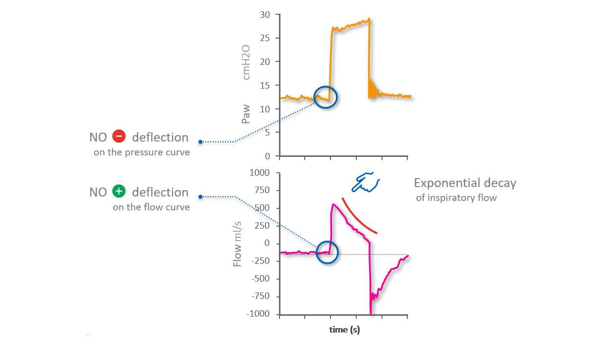
An auto-triggered breath is one triggered by the ventilator without an inspiratory effort made by the patient. Auto-triggering can be identified by watching both the Flow and Paw waveforms. There is no sign of the patient’s inspiratory effort, with neither a clear negative deflection of Paw, nor a clear positive deflection of Flow (with the exception of minimal oscillations due to cardiac activity (see Figure 5 below).

However, the strongest indication of auto-triggering can be seen on the inspiratory Flow waveform, which shows a very early peak followed by exponential decay: The whole inspiration is due to passive inflation. The shape of the peak itself is also an indicator: in the case of passive inspiration, the peak is a spike, whereas during synchronized inspiration it is more curved.

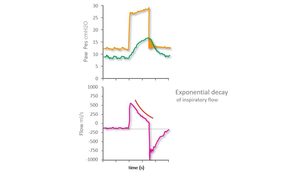
As on the Flow and Paw waveforms, there is no negative deflection visible on the Pes reference waveform that would normally indicate the start of the inspiratory effort (see Figure 6 below).

Flow and pressure waveforms showing no patient activity
Figure 5 (Image modified from Mojoli et al. Critical Care (2022) 26:32)
Flow and pressure waveforms showing no patient activity
Figure 5 (Image modified from Mojoli et al. Critical Care (2022) 26:32)
Pes waveform showing no negative deflection
Figure 6 (Image modified from Mojoli et al. Critical Care (2022) 26:32)
Pes waveform showing no negative deflection
Figure 6 (Image modified from Mojoli et al. Critical Care (2022) 26:32)

Major asynchrony - Ineffective inspiratory effort

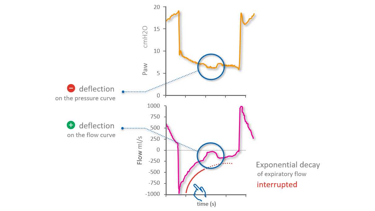
An ineffective inspiratory effort is an inspiratory effort made by the patient during the ventilator’s expiratory phase and not recognized – and therefore not supported - by the ventilator.

On the Flow waveform, you can identify the ineffective effort as a positive deflection temporarily interrupting the normal exponential decay of flow. On the Paw waveform, a  minimal negative deflection occurs simultaneously (see Figure 7 below).

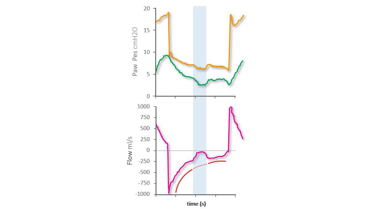
On the Pes reference waveform, you can see the beginning and end of the ineffective inspiratory effort shown as a negative deflection of Pes interrupting the normal expiratory decrease in Pes  (see Figure 8 below).

Flow and pressure waveforms with simultaneous activity
Figure 7 (Image modified from Mojoli et al. Critical Care (2022) 26:32)
Flow and pressure waveforms with simultaneous activity
Figure 7 (Image modified from Mojoli et al. Critical Care (2022) 26:32)
Pes waveform showing interruption to normal expiratory decrease
Figure 8 (Image modified from Mojoli et al. Critical Care (2022) 26:32)
Pes waveform showing interruption to normal expiratory decrease
Figure 8 (Image modified from Mojoli et al. Critical Care (2022) 26:32)
Asynchronies reference card

Learn to spot common asynchronies! Free reference card

Our asynchrony reference card gives you an overview of the most common asynchrony types, their causes, and how to detect them.

Timing of inspiratory muscle activity detected from airway pressure and flow during pressure support ventilation: the waveform method.

Mojoli F, Pozzi M, Orlando A, et al. Timing of inspiratory muscle activity detected from airway pressure and flow during pressure support ventilation: the waveform method. Crit Care. 2022;26(1):32. Published 2022 Jan 30. doi:10.1186/s13054-022-03895-4


BACKGROUND

Whether respiratory efforts and their timing can be reliably detected during pressure support ventilation using standard ventilator waveforms is unclear. This would give the opportunity to assess and improve patient-ventilator interaction without the need of special equipment.

METHODS

In 16 patients under invasive pressure support ventilation, flow and pressure waveforms were obtained from proximal sensors and analyzed by three trained physicians and one resident to assess patient's spontaneous activity. A systematic method (the waveform method) based on explicit rules was adopted. Esophageal pressure tracings were analyzed independently and used as reference. Breaths were classified as assisted or auto-triggered, double-triggered or ineffective. For assisted breaths, trigger delay, early and late cycling (minor asynchronies) were diagnosed. The percentage of breaths with major asynchronies (asynchrony index) and total asynchrony time were computed.

RESULTS

Out of 4426 analyzed breaths, 94.1% (70.4-99.4) were assisted, 0.0% (0.0-0.2) auto-triggered and 5.8% (0.4-29.6) ineffective. Asynchrony index was 5.9% (0.6-29.6). Total asynchrony time represented 22.4% (16.3-30.1) of recording time and was mainly due to minor asynchronies. Applying the waveform method resulted in an inter-operator agreement of 0.99 (0.98-0.99); 99.5% of efforts were detected on waveforms and agreement with the reference in detecting major asynchronies was 0.99 (0.98-0.99). Timing of respiratory efforts was accurately detected on waveforms: AUC for trigger delay, cycling delay and early cycling was 0.865 (0.853-0.876), 0.903 (0.892-0.914) and 0.983 (0.970-0.991), respectively.

CONCLUSIONS

Ventilator waveforms can be used alone to reliably assess patient's spontaneous activity and patient-ventilator interaction provided that a systematic method is adopted.

Related articles. Get a deeper look