Back

片肺換気に対する肺保護換気アプローチ

Article

Author: Munir Karjaghli, Clinical Applications Specialist, Hamilton Medical AG

Date of first publication: 15.11.2021

急性呼吸窮迫症候群(ARDS)と急性肺傷害(ALI)は大きな肺切除の後に認められる合併症で、しばしば命に関わります。大規模コホート研究では、大きな肺切除後のARDSおよびALIの発生率は2%~4%と報告されています(1-3)。これらの合併症が発生したときの死亡率は50%~70%です(1-3)。
片肺換気に対する肺保護換気アプローチ

要点

  • 急性呼吸窮迫症候群(ARDS)と急性肺傷害(ALI)は大きな肺切除の後に認められる合併症で、しばしば命に関わります。
  • 胸部手術中および術後の片肺換気は、容量損傷、圧損傷、無気肺損傷、酸素中毒のリスクを高めます。
  • 片肺換気中に許容性高炭酸ガス血症、一回換気量の低減、呼気終末陽圧の増加、人工呼吸器圧力の制限、リクルートメント手技などの肺保護換気プロトコルを実施すると、急性肺傷害のリスクが低下する可能性があります。

肺傷害の原因

片肺換気中の人工呼吸には3つの目標があります。それは、(I)二酸化炭素除去を助ける、(II)酸素化を維持する、(III)術後の肺機能不全を低減する、です。片肺換気中の最適な人工呼吸戦略を決定するためにさまざまな研究が行われています。

周術期ALIには多くの異なる要因が寄与する可能性があります。肺傷害は、炎症性または生化学的要因に加えて、過拡張、過灌流、周期的リクルートメント/デリクルートメントによる機械的応力にも起因します。胸部手術を受けた患者では、「マルチヒット」理論により、手術関連要因、片肺換気、基礎疾患および併存疾患、過去の治療、その他の不特定事象の組み合わせによってALIの起こりやすさが高まる可能性があることが示唆されています(Lytle FT, Brown DR. Appropriate ventilatory settings for thoracic surgery: intraoperative and postoperative. Semin Cardiothorac Vasc Anesth. 2008;12(2):97-108. doi:10.1177/10892532083198694​)。

胸部手術中および術後の片肺換気は、容量損傷、圧損傷、無気肺損傷、酸素中毒のリスクを高めます。これらはすべて、人工呼吸器誘発肺傷害を引き起こす重篤な合併症です(Lohser J. Evidence-based management of one-lung ventilation. Anesthesiol Clin. 2008;26(2):241-v. doi:10.1016/j.anclin.2008.01.0115​)。

保護的片肺換気とは

片肺換気の管理について、臨床転帰の観点からある特定のアプローチを特に支持するデータはほとんどありません。何を保護的片肺換気とみなすかという定義は、主に専門家の意見、一般手術患者での両肺換気から収集されたエビデンス、および少数の臨床試験の影響を受けます。たとえば、一回換気量を片肺換気中の肺傷害に寄与する単一の要因として特定することは非常に困難です。今日まで、片肺換気中に呼気終末陽圧(PEEP)(Blank RS, Colquhoun DA, Durieux ME, et al. Management of One-lung Ventilation: Impact of Tidal Volume on Complications after Thoracic Surgery. Anesthesiology. 2016;124(6):1286-1295. doi:10.1097/ALN.00000000000011006​)、気道内圧制限、リクルートメント手技などの他の換気戦略なしで低一回換気量(VT)換気を実施した場合の具体的な利点を明確に示した研究はありません。禁忌がある場合を除き、これらの換気アプローチは、すべての胸部手術患者で術中と術後のどちらにも使用できます。リクルートメントとPEEPは、液体、炎症、麻酔剤、その他の未知の変数とともに、肺保護換気に寄与します(Slinger PD. Do Low Tidal Volumes Decrease Lung Injury During One-Lung Ventilation?. J Cardiothorac Vasc Anesth. 2017;31(5):1774-1775. doi:10.1053/j.jvca.2017.07.0057​)。十分なPEEPがなければ、片肺換気中の低VTは無気肺につながる可能性があり、その結果死亡リスクが増加します(Levin MA, McCormick PJ, Lin HM, Hosseinian L, Fischer GW. Low intraoperative tidal volume ventilation with minimal PEEP is associated with increased mortality. Br J Anaesth. 2014;113(1):97-108. doi:10.1093/bja/aeu0548​)。低いPEEPは、肺胞の安定化、肺胞の負担の軽減、無気肺の防止には不十分です。無気肺は麻酔をかけられたすべての手術患者の懸念事項ですが、片肺換気中には、より高い吸入酸素濃度が使用されること(吸収性無気肺)、および下側肺圧迫のリスクが増加すること(圧迫性無気肺)から、無気肺がより重篤になる可能性があります(Duggan M, Kavanagh BP. Pulmonary atelectasis: a pathogenic perioperative entity. Anesthesiology. 2005;102(4):838-854. doi:10.1097/00000542-200504000-000219​)。

一回換気量よりもドライビングプレッシャー?

肺がん手術において片肺換気中にVTの低減、PEEPの増加、人工呼吸器圧力の制限、リクルートメント手技などの肺保護換気プロトコルを実施した後に行われたある後ろ向き研究では、急性肺傷害のリスクが低かったことがわかりました(Licker M, Diaper J, Villiger Y, et al. Impact of intraoperative lung-protective interventions in patients undergoing lung cancer surgery. Crit Care. 2009;13(2):R41. doi:10.1186/cc776210​)。VTの厳密な影響は不明ですが、新しいエビデンスは、VTまたはPEEPではなく気道ドライビングプレッシャーが術後肺合併症リスクの潜在的予測因子であることを示唆しており、胸部手術における片肺換気中のドライビングプレッシャー誘導換気の実施は、従来の肺保護換気と比較して術後肺合併症の発生率が低いことと関連していました(Park M, Ahn HJ, Kim JA, et al. Driving Pressure during Thoracic Surgery: A Randomized Clinical Trial. Anesthesiology. 2019;130(3):385-393. doi:10.1097/ALN.000000000000260011​)。

片肺換気中の人工呼吸管理に関する実践ガイドライン

Society for Translational Medicineが発行した肺葉切除を受ける患者の人工呼吸管理に関する臨床実践ガイドラインでは、片肺換気の現在のエビデンスに基づいて推奨事項が提案されています(Gao S, Zhang Z, Brunelli A, et al. The Society for Translational Medicine: clinical practice guidelines for mechanical ventilation management for patients undergoing lobectomy. J Thorac Dis. 2017;9(9):3246-3254. doi:10.21037/jtd.2017.08.16612​)。

  • 許容性/治療的高炭酸ガス血症:肺葉切除手術中に片肺換気を受ける患者では、二酸化炭素分圧を50~70 mmHgに維持すると有益である可能性がある。
  • 一回換気量4~6 mL/kgとPEEP 5~8 hPaを使用し、ドライビングプレッシャーを15 hPa未満に維持する肺保護換気は、現在のエビデンスに基づくと合理的なように思われる。
  • 肺胞リクルートメント(オープンラング換気)は、片肺換気で肺葉切除手術を受ける患者に有益である可能性がある。
  • プレッシャーコントロール換気(CMV-PC)またはプレッシャーコントロールボリューム保証換気(CMV-vtPC)はボリュームコントロール換気(CMV-VC)よりも推奨され、片肺換気で肺切除を受ける患者で使用できる。
  • 十分な動脈血酸素飽和度を維持するために必要な最低のFiO2を適用することは合理的である。
  • I:E比が1:1以上のコントロールされた人工呼吸は、片肺換気を受ける患者において合理的である。

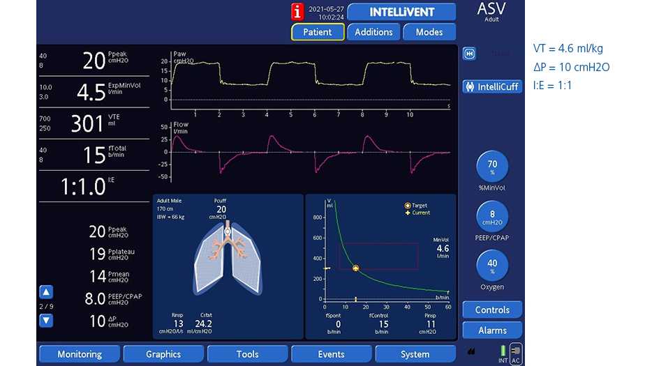
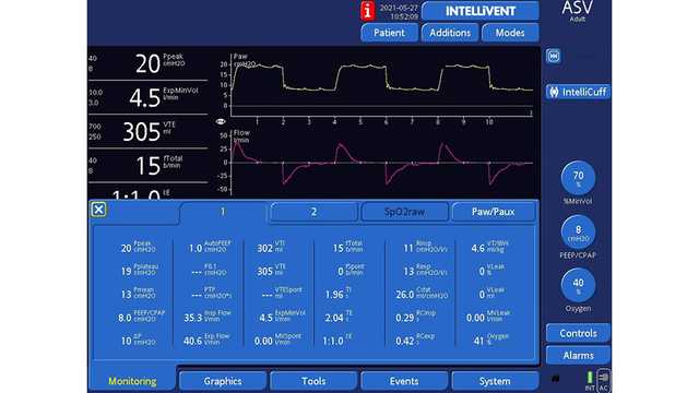
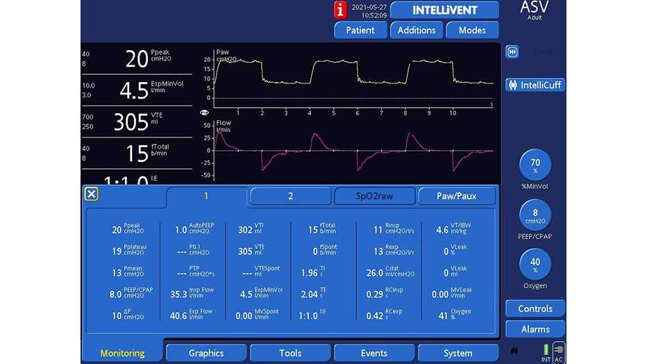
すべてのHamilton Medical人工呼吸器で使用可能なアダプティブサポートベンチレーション®(ASV®)モードは、片肺換気で推奨される一回換気量とドライビングプレッシャーに準拠した肺保護戦略を自動的に実行します。さらに、完全にクローズドループモードのINTELLiVENT®-ASV (Not available in all marketsA​) には、許容性高炭酸ガス血症を自動的に実現し、十分な動脈血酸素飽和度を維持するために必要な最低のFiO2を適用するオプションを提供します。

Weilerらの研究によると、ASVは、片肺換気の状況が非常に変わりやすい場合でも患者を安全に換気できます(Weiler N, Eberle B, Heinrichs W. Adaptive lung ventilation (ALV) during anesthesia for pulmonary surgery: automatic response to transitions to and from one-lung ventilation. J Clin Monit Comput. 1998;14(4):245-252. doi:10.1023/a:100997482523713​)。

図1と2は、ASVで換気を受けながら右肺全摘術を受けた61歳の男性患者を示しています。

参考文献は下記をご参照ください。(Ruffini E, Parola A, Papalia E, et al. Frequency and mortality of acute lung injury and acute respiratory distress syndrome after pulmonary resection for bronchogenic carcinoma. Eur J Cardiothorac Surg. 2001;20(1):30-37. doi:10.1016/s1010-7940(01)00760-61​, Kutlu CA, Williams EA, Evans TW, Pastorino U, Goldstraw P. Acute lung injury and acute respiratory distress syndrome after pulmonary resection. Ann Thorac Surg. 2000;69(2):376-380. doi:10.1016/s0003-4975(99)01090-52​, Shapiro M, Swanson SJ, Wright CD, et al. Predictors of major morbidity and mortality after pneumonectomy utilizing the Society for Thoracic Surgeons General Thoracic Surgery Database. Ann Thorac Surg. 2010;90(3):927-935. doi:10.1016/j.athoracsur.2010.05.0413​)

Display showing dynamic lung and ASV graph
図版 1
Display showing dynamic lung and ASV graph
図版 1
Display showing dynamic lung and ASV graph
図版 2
Display showing dynamic lung and ASV graph
図版 2

Footnotes

  • A. Not available in all markets

References

  1. 1. Ruffini E, Parola A, Papalia E, et al. Frequency and mortality of acute lung injury and acute respiratory distress syndrome after pulmonary resection for bronchogenic carcinoma. Eur J Cardiothorac Surg. 2001;20(1):30-37. doi:10.1016/s1010-7940(01)00760-6
  2. 2. Kutlu CA, Williams EA, Evans TW, Pastorino U, Goldstraw P. Acute lung injury and acute respiratory distress syndrome after pulmonary resection. Ann Thorac Surg. 2000;69(2):376-380. doi:10.1016/s0003-4975(99)01090-5
  3. 3. Shapiro M, Swanson SJ, Wright CD, et al. Predictors of major morbidity and mortality after pneumonectomy utilizing the Society for Thoracic Surgeons General Thoracic Surgery Database. Ann Thorac Surg. 2010;90(3):927-935. doi:10.1016/j.athoracsur.2010.05.041
  4. 4. Lytle FT, Brown DR. Appropriate ventilatory settings for thoracic surgery: intraoperative and postoperative. Semin Cardiothorac Vasc Anesth. 2008;12(2):97-108. doi:10.1177/1089253208319869
  5. 5. Lohser J. Evidence-based management of one-lung ventilation. Anesthesiol Clin. 2008;26(2):241-v. doi:10.1016/j.anclin.2008.01.011
  6. 6. Blank RS, Colquhoun DA, Durieux ME, et al. Management of One-lung Ventilation: Impact of Tidal Volume on Complications after Thoracic Surgery. Anesthesiology. 2016;124(6):1286-1295. doi:10.1097/ALN.0000000000001100
  7. 7. Slinger PD. Do Low Tidal Volumes Decrease Lung Injury During One-Lung Ventilation?. J Cardiothorac Vasc Anesth. 2017;31(5):1774-1775. doi:10.1053/j.jvca.2017.07.005
  8. 8. Levin MA, McCormick PJ, Lin HM, Hosseinian L, Fischer GW. Low intraoperative tidal volume ventilation with minimal PEEP is associated with increased mortality. Br J Anaesth. 2014;113(1):97-108. doi:10.1093/bja/aeu054
  9. 9. Duggan M, Kavanagh BP. Pulmonary atelectasis: a pathogenic perioperative entity. Anesthesiology. 2005;102(4):838-854. doi:10.1097/00000542-200504000-00021
  10. 10. Licker M, Diaper J, Villiger Y, et al. Impact of intraoperative lung-protective interventions in patients undergoing lung cancer surgery. Crit Care. 2009;13(2):R41. doi:10.1186/cc7762
  11. 11. Park M, Ahn HJ, Kim JA, et al. Driving Pressure during Thoracic Surgery: A Randomized Clinical Trial. Anesthesiology. 2019;130(3):385-393. doi:10.1097/ALN.0000000000002600
  12. 12. Gao S, Zhang Z, Brunelli A, et al. The Society for Translational Medicine: clinical practice guidelines for mechanical ventilation management for patients undergoing lobectomy. J Thorac Dis. 2017;9(9):3246-3254. doi:10.21037/jtd.2017.08.166
  13. 13. Weiler N, Eberle B, Heinrichs W. Adaptive lung ventilation (ALV) during anesthesia for pulmonary surgery: automatic response to transitions to and from one-lung ventilation. J Clin Monit Comput. 1998;14(4):245-252. doi:10.1023/a:1009974825237

Frequency and mortality of acute lung injury and acute respiratory distress syndrome after pulmonary resection for bronchogenic carcinoma.

Ruffini E, Parola A, Papalia E, et al. Frequency and mortality of acute lung injury and acute respiratory distress syndrome after pulmonary resection for bronchogenic carcinoma. Eur J Cardiothorac Surg. 2001;20(1):30-37. doi:10.1016/s1010-7940(01)00760-6

OBJECTIVE We reviewed the frequency and mortality of acute lung injury (ALI) and acute respiratory distress syndrome (ARDS) in our population of patients submitted to pulmonary resection for primary bronchogenic carcinoma. METHODS From January 1993 to December 1999, a total of 1221 patients received pulmonary resection for primary bronchogenic carcinoma. Of these, 27 met the criteria of post-operative ALI/ARDS. There were 24 men and three women with a mean age of 64 years (range 45--79). Pre-operatively, predicted mean of PaO(2), PaCO(2) and %FEV1 were 72 mmHg (57--86), 37 mmHg (33--42) and 80% (37--114), respectively. Associated cardiac risk factors were present in eight patients. Three patients (11%) had pre-operative radiotherapy. Surgical-pathologic staging included 14 patients at Stage I, 8 patients at Stage II, four patients at Stage IIIa and one patient at Stage IIIb. RESULTS ALI/ARDS occurred in 2.2% of our operated lung cancer patients. ALI was diagnosed in 10 patients and ARDS in 17 patients. The mean time of presentation following surgery was 4 days (range 1--10) and 6 days (1--13) for ALI and ARDS, respectively. According to the type of operation, the frequency was highest following right pneumonectomy (4.5%), followed by sublobar resection (3.2%), left pneumonectomy (3%), bilobectomy (2.4%), and lobectomy (2%). The frequency following extended operations was 4%. No differences were found between the ALI/ARDS group and the total population of resected lung cancer patients (control group) with respect to sex, mean age, pre-operative blood gases, %FEV1, surgical--pathologic staging and the use of pre-operative radiotherapy. Four patients with ALI (40%) and 10 patients with ARDS (59%) died. Mortality was highest following right pneumonectomy, extended operations and sublobar resections. Hospital mortality of the total population of operated lung cancer patients in the same period was 2.8% (34 patients). ALI/ARDS accounted for 41% of our hospital mortality. CONCLUSIONS (1) ALI/ARDS is a severe complication following resection for primary bronchogenic carcinoma. (2) We did not detect any significant difference between the ALI/ARDS group and the control group regarding age, pre-operative lung function, staging and pre-operative radiotherapy. (3) ALI/ARDS is associated with high mortality, the highest mortality rates having been observed following right pneumonectomy and extended operation; it currently represents our leading cause of death following pulmonary resection for lung carcinoma. (4) ALI/ARDS may also occur after sublobar resections with an associated high mortality rate.

Acute lung injury and acute respiratory distress syndrome after pulmonary resection.

Kutlu CA, Williams EA, Evans TW, Pastorino U, Goldstraw P. Acute lung injury and acute respiratory distress syndrome after pulmonary resection. Ann Thorac Surg. 2000;69(2):376-380. doi:10.1016/s0003-4975(99)01090-5

BACKGROUND In this study we investigate the frequency and mortality of acute lung injury (ALI) and acute respiratory distress syndrome (ARDS) after pulmonary resection. METHODS Patients that underwent pulmonary resection at the Royal Brompton Hospital between 1991 and 1997 were included. The case notes of all patients developing postoperative complications were retrospectively reviewed. RESULTS The overall combined frequency of ALI and ARDS was 3.9%. The frequency was higher in patients over 60 years of age, males and those undergoing resection for lung cancer. ALI/ARDS caused 72.5% of the total mortality after resection in this series. CONCLUSIONS In our experience ALI and ARDS are major causes of mortality after lung resection.

Predictors of major morbidity and mortality after pneumonectomy utilizing the Society for Thoracic Surgeons General Thoracic Surgery Database.

Shapiro M, Swanson SJ, Wright CD, et al. Predictors of major morbidity and mortality after pneumonectomy utilizing the Society for Thoracic Surgeons General Thoracic Surgery Database. Ann Thorac Surg. 2010;90(3):927-935. doi:10.1016/j.athoracsur.2010.05.041

BACKGROUND Pneumonectomy is associated with a significant incidence of perioperative morbidity and mortality. The purpose of this study is to identify the risk factors responsible for adverse outcomes in patients after pneumonectomy utilizing The Society of Thoracic Surgeons General Thoracic Surgery Database (STS GTDB). METHODS All patients who had undergone pneumonectomy between January 2002 and December 2007 were identified in the STS GTDB. Among 80 participating centers, 1,267 patients were selected. Logistic regression analysis was performed on preoperative variables for major adverse outcomes. RESULTS The rate of major adverse perioperative events was 30.4%, including 71 patients who died (5.6%). Major morbidity was defined as pneumonia, adult respiratory distress syndrome, empyema, sepsis, bronchopleural fistula, pulmonary embolism, ventilatory support beyond 48 hours, reintubation, tracheostomy, atrial or ventricular arrhythmias requiring treatment, myocardial infarct, reoperation for bleeding, and central neurologic event. Patients with major morbidity had a longer mean length of stay compared with patients without major morbidity (13.3 versus 6.1 days, p < 0.001). Independent predictors of major adverse outcomes were age 65 years or older (p < 0.001), male sex (p = 0.026), congestive heart failure (p = 0.04), forced expiratory volume in 1 second less than 60% of predicted (p = 0.01), benign lung disease (p = 0.006), and requiring extrapleural pneumonectomy (p = 0.018). Among patients with lung carcinoma, those receiving neoadjuvant chemoradiotherapy were more at risk for major morbidity than patients without induction therapy (p = 0.049). CONCLUSIONS The mortality rate after pneumonectomy by thoracic surgeons participating in the STS database compares favorably to that in previously published studies. We identified risk factors for major adverse outcomes in patients undergoing pneumonectomy.

Appropriate ventilatory settings for thoracic surgery: intraoperative and postoperative.

Lytle FT, Brown DR. Appropriate ventilatory settings for thoracic surgery: intraoperative and postoperative. Semin Cardiothorac Vasc Anesth. 2008;12(2):97-108. doi:10.1177/1089253208319869

Mechanical ventilation of patients undergoing thoracic surgery is often challenging. These patients frequently have significant underlying comorbidities, including cardiopulmonary disease, and often must undergo 1-lung ventilation. Perioperative respiratory complications are common and are multifactorial in etiology. Increasing evidence suggests that mechanical ventilation is associated with, and may even cause, lung damage in both sick and healthy patients. Gas exchange to provide acceptable end-organ oxygenation remains a primary goal but so too is minimization of risks for acute lung injury. Every ventilator strategy is associated with potential beneficial and adverse side effects. Understanding the impact of various ventilation strategies allows clinicians to provide optimal care for patients.

Evidence-based management of one-lung ventilation.

Lohser J. Evidence-based management of one-lung ventilation. Anesthesiol Clin. 2008;26(2):241-v. doi:10.1016/j.anclin.2008.01.011

One-lung ventilation (OLV) is essential for many thoracic and an increasing number of non-thoracic minimally invasive procedures. Beyond the well-recognized disturbance of ventilation-perfusion matching, recent years have seen a mounting body of evidence implicating OLV in the creation of acute lung injury. After reviewing the fundamentals of OLV physiology, this article examines the evidence for altering individual ventilatory parameters toward protective OLV.

Management of One-lung Ventilation: Impact of Tidal Volume on Complications after Thoracic Surgery.

Blank RS, Colquhoun DA, Durieux ME, et al. Management of One-lung Ventilation: Impact of Tidal Volume on Complications after Thoracic Surgery. Anesthesiology. 2016;124(6):1286-1295. doi:10.1097/ALN.0000000000001100

BACKGROUND The use of lung-protective ventilation (LPV) strategies may minimize iatrogenic lung injury in surgical patients. However, the identification of an ideal LPV strategy, particularly during one-lung ventilation (OLV), remains elusive. This study examines the role of ventilator management during OLV and its impact on clinical outcomes. METHODS Data were retrospectively collected from the hospital electronic medical record and the Society of Thoracic Surgery database for subjects undergoing thoracic surgery with OLV between 2012 and 2014. Mean tidal volume (VT) during two-lung ventilation and OLV and ventilator driving pressure (ΔP) (plateau pressure - positive end-expiratory pressure [PEEP]) were analyzed for the 1,019 cases that met the inclusion criteria. Associations between ventilator parameters and clinical outcomes were examined by multivariate linear regression. RESULTS After the initiation of OLV, 73.3, 43.3, 18.8, and 7.2% of patients received VT greater than 5, 6, 7, and 8 ml/kg predicted body weight, respectively. One hundred and eighty-four primary and 288 secondary outcome events were recorded. In multivariate logistic regression modeling, VT was inversely related to the incidence of respiratory complications (odds ratio, 0.837; 95% CI, 0.729 to 0.958), while ΔP predicted the development of major morbidity when modeled with VT (odds ratio, 1.034; 95% CI, 1.001 to 1.068). CONCLUSIONS Low VT per se (i.e., in the absence of sufficient PEEP) has not been unambiguously demonstrated to be beneficial. The authors found that a large proportion of patients continue to receive high VT during OLV and that VT was inversely related to the incidence of respiratory complications and major postoperative morbidity. While low (physiologically appropriate) VT is an important component of an LPV strategy for surgical patients during OLV, current evidence suggests that, without adequate PEEP, low VT does not prevent postoperative respiratory complications. Thus, use of physiologic VT may represent a necessary, but not independently sufficient, component of LPV.

Do Low Tidal Volumes Decrease Lung Injury During One-Lung Ventilation?

Slinger PD. Do Low Tidal Volumes Decrease Lung Injury During One-Lung Ventilation?. J Cardiothorac Vasc Anesth. 2017;31(5):1774-1775. doi:10.1053/j.jvca.2017.07.005

Low intraoperative tidal volume ventilation with minimal PEEP is associated with increased mortality.

Levin MA, McCormick PJ, Lin HM, Hosseinian L, Fischer GW. Low intraoperative tidal volume ventilation with minimal PEEP is associated with increased mortality. Br J Anaesth. 2014;113(1):97-108. doi:10.1093/bja/aeu054

BACKGROUND Anaesthetists have traditionally ventilated patients' lungs with tidal volumes (TVs) between 10 and 15 ml kg(-1) of ideal body weight (IBW), without the use of PEEP. Over the past decade, influenced by the results of the Acute Respiratory Distress Syndrome Network trial, many anaesthetists have begun using lower TVs during surgery. It is unclear whether the benefits of low TV ventilation can be extended into the perioperative period. METHODS We reviewed the records of 29 343 patients who underwent general anaesthesia with mechanical ventilation between January 1, 2008 and December 31, 2011. We calculated TV kg(-1) IBW, PEEP, peak inspiratory pressure (PIP), and dynamic compliance. Cox regression analysis with propensity score matching was performed to examine the association between TV and 30-day mortality. RESULTS Median TV was 8.6 [7.7-9.6] ml kg(-1) IBW with minimal PEEP [4.0 (2.2-5.0) cm H2O]. A significant reduction in TV occurred over the study period, from 9 ml kg(-1) IBW in 2008 to 8.3 ml kg(-1) IBW in 2011 (P=0.01). Low TV 6-8 ml kg(-1) IBW was associated with a significant increase in 30-day mortality vs TV 8-10 ml kg(-1) IBW: hazard ratio (HR) 1.6 [95% confidence interval (CI) [1.25-2.08], P=0.0002]. The association remained significant after matching: HR 1.63 [95% CI (1.22-2.18), P<0.001]. There was only a weak correlation between TV kg(-1) IBW and dynamic compliance (r=-0.006, P=0.31) and a weak-to-moderate correlation between TV kg(-1) IBW and PIP (r=0.32 P<0.0001). CONCLUSIONS Use of low intraoperative TV with minimal PEEP is associated with an increased risk of 30-day mortality.

Pulmonary atelectasis: a pathogenic perioperative entity.

Duggan M, Kavanagh BP. Pulmonary atelectasis: a pathogenic perioperative entity. Anesthesiology. 2005;102(4):838-854. doi:10.1097/00000542-200504000-00021

Atelectasis occurs in the dependent parts of the lungs of most patients who are anesthetized. Development of atelectasis is associated with decreased lung compliance, impairment of oxygenation, increased pulmonary vascular resistance, and development of lung injury. The adverse effects of atelectasis persist into the postoperative period and can impact patient recovery. This review article focuses on the causes, nature, and diagnosis of atelectasis. The authors discuss the effects and implications of atelectasis in the perioperative period and illustrate how preventive measures may impact outcome. In addition, they examine the impact of atelectasis and its prevention in acute lung injury.

Impact of intraoperative lung-protective interventions in patients undergoing lung cancer surgery.

Licker M, Diaper J, Villiger Y, et al. Impact of intraoperative lung-protective interventions in patients undergoing lung cancer surgery. Crit Care. 2009;13(2):R41. doi:10.1186/cc7762

INTRODUCTION In lung cancer surgery, large tidal volume and elevated inspiratory pressure are known risk factors of acute lung (ALI). Mechanical ventilation with low tidal volume has been shown to attenuate lung injuries in critically ill patients. In the current study, we assessed the impact of a protective lung ventilation (PLV) protocol in patients undergoing lung cancer resection. METHODS We performed a secondary analysis of an observational cohort. Demographic, surgical, clinical and outcome data were prospectively collected over a 10-year period. The PLV protocol consisted of small tidal volume, limiting maximal pressure ventilation and adding end-expiratory positive pressure along with recruitment maneuvers. Multivariate analysis with logistic regression was performed and data were compared before and after implementation of the PLV protocol: from 1998 to 2003 (historical group, n = 533) and from 2003 to 2008 (protocol group, n = 558). RESULTS Baseline patient characteristics were similar in the two cohorts, except for a higher cardiovascular risk profile in the intervention group. During one-lung ventilation, protocol-managed patients had lower tidal volume (5.3 +/- 1.1 vs. 7.1 +/- 1.2 ml/kg in historical controls, P = 0.013) and higher dynamic compliance (45 +/- 8 vs. 32 +/- 7 ml/cmH2O, P = 0.011). After implementing PLV, there was a decreased incidence of acute lung injury (from 3.7% to 0.9%, P < 0.01) and atelectasis (from 8.8 to 5.0, P = 0.018), fewer admissions to the intensive care unit (from 9.4% vs. 2.5%, P < 0.001) and shorter hospital stay (from 14.5 +/- 3.3 vs. 11.8 +/- 4.1, P < 0.01). When adjusted for baseline characteristics, implementation of the open-lung protocol was associated with a reduced risk of acute lung injury (adjusted odds ratio of 0.34 with 95% confidence interval of 0.23 to 0.75; P = 0.002). CONCLUSIONS Implementing an intraoperative PLV protocol in patients undergoing lung cancer resection was associated with improved postoperative respiratory outcomes as evidence by significantly reduced incidences of acute lung injury and atelectasis along with reduced utilization of intensive care unit resources.

Driving Pressure during Thoracic Surgery: A Randomized Clinical Trial.

Park M, Ahn HJ, Kim JA, et al. Driving Pressure during Thoracic Surgery: A Randomized Clinical Trial. Anesthesiology. 2019;130(3):385-393. doi:10.1097/ALN.0000000000002600

WHAT WE ALREADY KNOW ABOUT THIS TOPIC Driving pressure (plateau minus end-expiratory airway pressure) is a target in patients with acute respiratory distress syndrome, and is proposed as a target during general anesthesia for patients with normal lungs. It has not been reported for thoracic anesthesia where isolated, inflated lungs may be especially at risk. WHAT THIS ARTICLE TELLS US THAT IS NEW In a double-blinded, randomized trial (292 patients), minimized driving pressure compared with standard protective ventilation was associated with less postoperative pneumonia or acute respiratory distress syndrome. BACKGROUND Recently, several retrospective studies have suggested that pulmonary complication is related with driving pressure more than any other ventilatory parameter. Thus, the authors compared driving pressure-guided ventilation with conventional protective ventilation in thoracic surgery, where lung protection is of the utmost importance. The authors hypothesized that driving pressure-guided ventilation decreases postoperative pulmonary complications more than conventional protective ventilation. METHODS In this double-blind, randomized, controlled study, 292 patients scheduled for elective thoracic surgery were included in the analysis. The protective ventilation group (n = 147) received conventional protective ventilation during one-lung ventilation: tidal volume 6 ml/kg of ideal body weight, positive end-expiratory pressure (PEEP) 5 cm H2O, and recruitment maneuver. The driving pressure group (n = 145) received the same tidal volume and recruitment, but with individualized PEEP which produces the lowest driving pressure (plateau pressure-PEEP) during one-lung ventilation. The primary outcome was postoperative pulmonary complications based on the Melbourne Group Scale (at least 4) until postoperative day 3. RESULTS Melbourne Group Scale of at least 4 occurred in 8 of 145 patients (5.5%) in the driving pressure group, as compared with 18 of 147 (12.2%) in the protective ventilation group (P = 0.047, odds ratio 0.42; 95% CI, 0.18 to 0.99). The number of patients who developed pneumonia or acute respiratory distress syndrome was less in the driving pressure group than in the protective ventilation group (10/145 [6.9%] vs. 22/147 [15.0%], P = 0.028, odds ratio 0.42; 95% CI, 0.19 to 0.92). CONCLUSIONS Application of driving pressure-guided ventilation during one-lung ventilation was associated with a lower incidence of postoperative pulmonary complications compared with conventional protective ventilation in thoracic surgery.

The Society for Translational Medicine: clinical practice guidelines for mechanical ventilation management for patients undergoing lobectomy.

Gao S, Zhang Z, Brunelli A, et al. The Society for Translational Medicine: clinical practice guidelines for mechanical ventilation management for patients undergoing lobectomy. J Thorac Dis. 2017;9(9):3246-3254. doi:10.21037/jtd.2017.08.166

Patients undergoing lobectomy are at significantly increased risk of lung injury. One-lung ventilation is the most commonly used technique to maintain ventilation and oxygenation during the operation. It is a challenge to choose an appropriate mechanical ventilation strategy to minimize the lung injury and other adverse clinical outcomes. In order to understand the available evidence, a systematic review was conducted including the following topics: (I) protective ventilation (PV); (II) mode of mechanical ventilation [e.g., volume controlled (VCV) versus pressure controlled (PCV)]; (III) use of therapeutic hypercapnia; (IV) use of alveolar recruitment (open-lung) strategy; (V) pre-and post-operative application of positive end expiratory pressure (PEEP); (VI) Inspired Oxygen concentration; (VII) Non-intubated thoracoscopic lobectomy; and (VIII) adjuvant pharmacologic options. The recommendations of class II are non-intubated thoracoscopic lobectomy may be an alternative to conventional one-lung ventilation in selected patients. The recommendations of class IIa are: (I) Therapeutic hypercapnia to maintain a partial pressure of carbon dioxide at 50-70 mmHg is reasonable for patients undergoing pulmonary lobectomy with one-lung ventilation; (II) PV with a tidal volume of 6 mL/kg and PEEP of 5 cmH2O are reasonable methods, based on current evidence; (III) alveolar recruitment [open lung ventilation (OLV)] may be beneficial in patients undergoing lobectomy with one-lung ventilation; (IV) PCV is recommended over VCV for patients undergoing lung resection; (V) pre- and post-operative CPAP can improve short-term oxygenation in patients undergoing lobectomy with one-lung ventilation; (VI) controlled mechanical ventilation with I:E ratio of 1:1 is reasonable in patients undergoing one-lung ventilation; (VII) use of lowest inspired oxygen concentration to maintain satisfactory arterial oxygen saturation is reasonable based on physiologic principles; (VIII) Adjuvant drugs such as nebulized budesonide, intravenous sivelestat and ulinastatin are reasonable and can be used to attenuate inflammatory response.

Adaptive lung ventilation (ALV) during anesthesia for pulmonary surgery: automatic response to transitions to and from one-lung ventilation.

Weiler N, Eberle B, Heinrichs W. Adaptive lung ventilation (ALV) during anesthesia for pulmonary surgery: automatic response to transitions to and from one-lung ventilation. J Clin Monit Comput. 1998;14(4):245-252. doi:10.1023/a:1009974825237

UNLABELLED Adaptive lung ventilation is a novel closed-loop-controlled ventilation system. Based upon instantaneous breath-to-breath analyses, the ALV controller adjusts ventilation patterns automatically to momentary respiratory mechanics. Its goal is to provide a preset alveolar ventilation (V'A) and, at the same time, minimize the work of breathing. Aims of our study were (1) to investigate changes in respiratory mechanics during transition to and from one-lung ventilation (OLV), (2) to describe the automated adaptation of the ventilatory pattern. METHODS With institutional approval and informed consent, 9 patients (33-72 y, 66-88 kg) underwent ALV during total intravenous anesthesia for pulmonary surgery. The ALV controller uses a pressure controlled ventilation mode. V'A is preset by the anesthesiologist. Flow, pressure, and CO2 are continuously measured at the DLT connector. The signals were read into a IBM compatible PC and processed using a linear one-compartment model of the lung to calculate breath-by-breath resistance (R), compliance (C), respiratory time constant (TC), serial dead space (VdS) and V'A. Based upon the results, the controller optimizes respiratory rate (RR) and tidal volume (VT) such as to achieve the preset V'A with the minimum work of breathing. In addition to V'A, only PEEP and FIO2 settings are at the anesthesiologist's discretion. All patients were ventilated using FIO2 = 1,0 and PEEP = 3 cm H2O. Parameters of respiratory mechanics, ventilation, and ABG were recorded during three 5-min periods: 10 min prior to OLV (1), 20 min after onset of OLV (II), and after chest closure (III). Data analyses used nonparametric comparisons of paired samples (Wilcoxon, Friedman) with Bonferroni's correction. Significance was assumed at p < 0.05. Values are given as medians (range). RESULTS 20 min after onset of OLV (II), resistance had approximately doubled compared with (1), compliance had decreased from 54 (36-81) to 50 (25-70) ml/cm H2O. TC remained stable at 1.4 (0.8-2.4) vs. 1.2 (0.9)-1.6) s. Institution of OLV was followed by a reproducible response of the ALV controller. The sudden changes in respiratory mechanics caused a transient reduction in VT by 42 (8-59)%, with RR unaffected. In order to reestablish the preset V'A, the controller increased inspiratory pressure in a stepwise fashion from 18 (14-23) to 27 (19-39) cm H2O, thereby increasing VT close to baseline (7.5 (6.6-9.0) ml/kg BW vs. 7.9 (5.4-11.7) ml/kg BW). The controller was, thus, effective in maintaining V'A. The minimum PaO2 during phase II was 101 mmHg. After chest closure, respiratory mechanics had returned to baseline. CONCLUSIONS Respiratory mechanics during transition to and from OLV are characterized by marked changes in R and C into opposite directions, leaving TC unaffected. The ALV controller manages these transitions successfully, and maintains V'A reliably without intervention by the anesthesiologist. VT during OLV was found to be consistently lower than recommended in the literature.