Back

補助換気中の患者でドライビングプレッシャーを測定する方法

Article

Author: Jean-Michel Arnal、サント・ミュス病院(フランス、トゥーロン)

Date of first publication: 18.09.2025

静的ドライビングプレッシャーをベッドサイドで体系的にモニタリングする必要がありますが、どのように測定すればよいのでしょうか?

静的ドライビングプレッシャーとは

補助換気モードを使用している人工呼吸器装着患者において、静的ドライビングプレッシャー(∆PSTAT)は単独でICU生存率と関連してしています(Bellani G, Grassi A, Sosio S, et al. Driving Pressure Is Associated with Outcome during Assisted Ventilation in Acute Respiratory Distress Syndrome. Anesthesiology. 2019;131(3):594-604. doi:10.1097/ALN.00000000000028461Grassi A, Bianchi I, Teggia Droghi M, et al. Increased Driving Pressure During Assisted Ventilation for Hypoxemic Respiratory Failure Is Associated with Lower ICU Survival: The ICEBERG Study. Am J Respir Crit Care Med. Published online June 20, 2025. doi:10.1164/rccm.202411-2146OC2)。したがって、安全な自発呼吸を確認し、自発呼吸誘発性肺傷害を防ぐため、臨床医はベッドサイドで∆PSTATを体系的にモニタリングする必要があります。∆PSTATは、呼吸中に呼吸器系にかかる弾性圧力の変化を表したもので、プラトー圧(PPLAT)からPEEPを差し引くことによって計算されます。

プラトー圧の取得方法

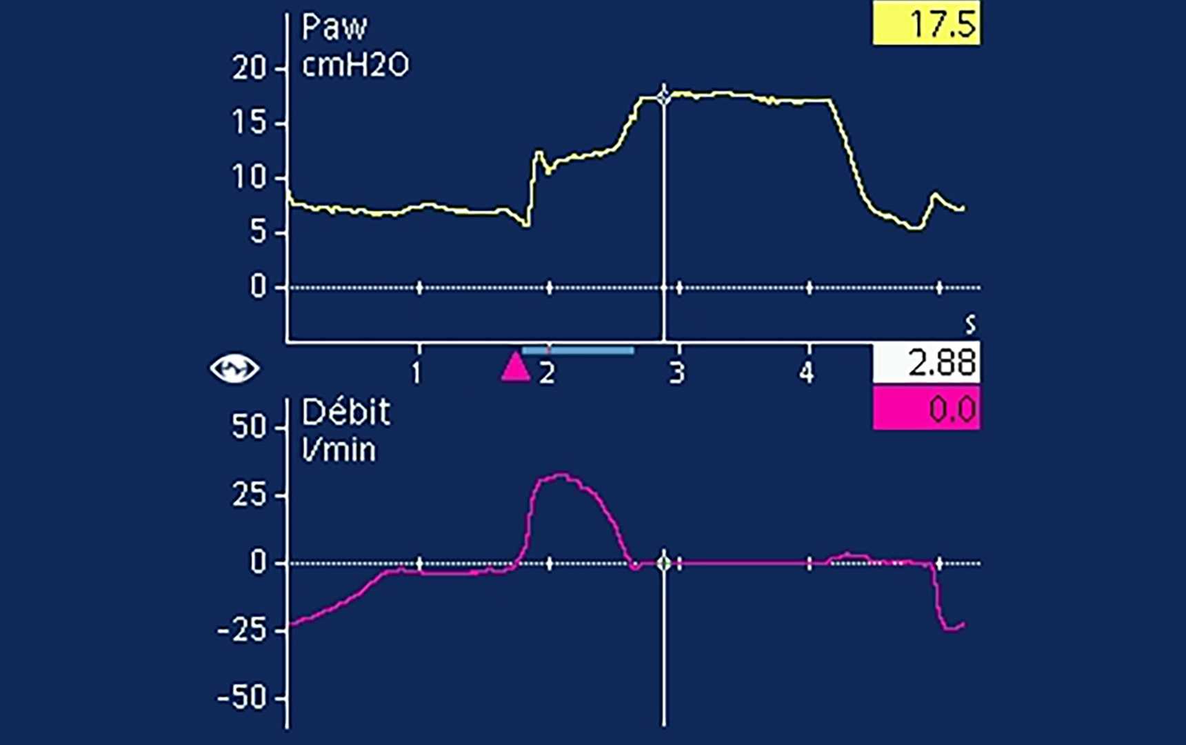
補助換気におけるPPLATの測定には吸気終末閉塞法を使用しますが(Foti G, Cereda M, Banfi G, Pelosi P, Fumagalli R, Pesenti A. End-inspiratory airway occlusion: a method to assess the pressure developed by inspiratory muscles in patients with acute lung injury undergoing pressure support. Am J Respir Crit Care Med. 1997;156(4 Pt 1):1210-1216. doi:10.1164/ajrccm.156.4.96-020313)、気道内圧のプラトーが視認できることが条件となります。補助換気の場合、吸気終末ホールドを行うと、気道内圧がピーク圧を超えて上昇し、その後プラトーになります(Bellani G, Grassi A, Sosio S, Foti G. Plateau and driving pressure in the presence of spontaneous breathing. Intensive Care Med. 2019;45(1):97-98. doi:10.1007/s00134-018-5311-94)。PPLATまでの気道内圧の上昇は、呼吸中の患者の吸気努力に対応しており、これをPmus指数(PMI)と呼びます(Foti G, Cereda M, Banfi G, Pelosi P, Fumagalli R, Pesenti A. End-inspiratory airway occlusion: a method to assess the pressure developed by inspiratory muscles in patients with acute lung injury undergoing pressure support. Am J Respir Crit Care Med. 1997;156(4 Pt 1):1210-1216. doi:10.1164/ajrccm.156.4.96-020313)。ホールドを開始すると気道内圧が急激に上昇し、その後すぐに平坦な部分が続きます。この部分がプラトー圧になります(図1)(Bianchi I, Grassi A, Pham T, et al. Reliability of plateau pressure during patient-triggered assisted ventilation. Analysis of a multicentre database. J Crit Care. 2022;68:96-103. doi:10.1016/j.jcrc.2021.12.0025)。観察研究では、PPLATはホールドの90%以上で読み取り可能であると報告されています(Bellani G, Grassi A, Sosio S, et al. Driving Pressure Is Associated with Outcome during Assisted Ventilation in Acute Respiratory Distress Syndrome. Anesthesiology. 2019;131(3):594-604. doi:10.1097/ALN.00000000000028461Bianchi I, Grassi A, Pham T, et al. Reliability of plateau pressure during patient-triggered assisted ventilation. Analysis of a multicentre database. J Crit Care. 2022;68:96-103. doi:10.1016/j.jcrc.2021.12.0025)。プラトーが視認できない場合、すなわちホールド中に吸気筋または呼気筋が活動した場合には、ホールドは読み取れません(図2)(Bellani G, Grassi A, Sosio S, et al. Driving Pressure Is Associated with Outcome during Assisted Ventilation in Acute Respiratory Distress Syndrome. Anesthesiology. 2019;131(3):594-604. doi:10.1097/ALN.00000000000028461)。

気道内圧波形とフロー波形を示すスクリーンショット。フロー波形でプラトーを視認できる
図1:17 cmH2OのPPLATでプラトーを視認できます。この患者では、PPEAKは12 cmH2Oで、PEEPは7 cmH2Oです。したがって、∆PSTATは10 cmH2O、PMIは5 cmH2Oになります。
気道内圧波形とフロー波形を示すスクリーンショット。フロー波形でプラトーを視認できる
図1:17 cmH2OのPPLATでプラトーを視認できます。この患者では、PPEAKは12 cmH2Oで、PEEPは7 cmH2Oです。したがって、∆PSTATは10 cmH2O、PMIは5 cmH2Oになります。
読み取り不能なプラトー圧を示すスクリーンショット
図2a:吸気筋の活動が原因でプラトー圧が読み取れない
読み取り不能なプラトー圧を示すスクリーンショット
図2a:吸気筋の活動が原因でプラトー圧が読み取れない
呼気筋の活動が原因で読み取り不能なプラトー圧を示すスクリーンショット
図2b:呼気筋の活動が原因でプラトー圧が読み取れない
呼気筋の活動が原因で読み取り不能なプラトー圧を示すスクリーンショット
図2b:呼気筋の活動が原因でプラトー圧が読み取れない
吸気筋と呼気筋の活動が原因で読み取り不能なプラトー圧を示すスクリーンショット
図2c:吸気筋と呼気筋の両方の活動が原因でプラトー圧が読み取れない
吸気筋と呼気筋の活動が原因で読み取り不能なプラトー圧を示すスクリーンショット
図2c:吸気筋と呼気筋の両方の活動が原因でプラトー圧が読み取れない

過大評価に注意する

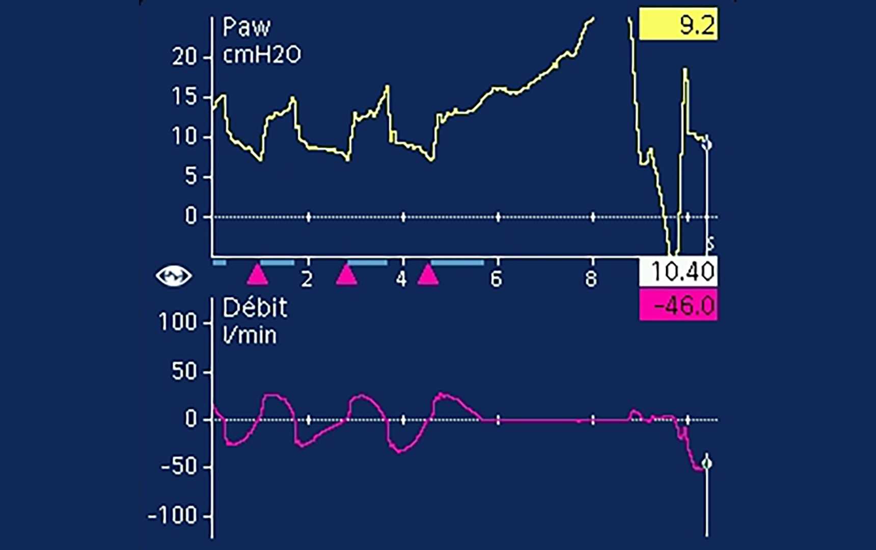
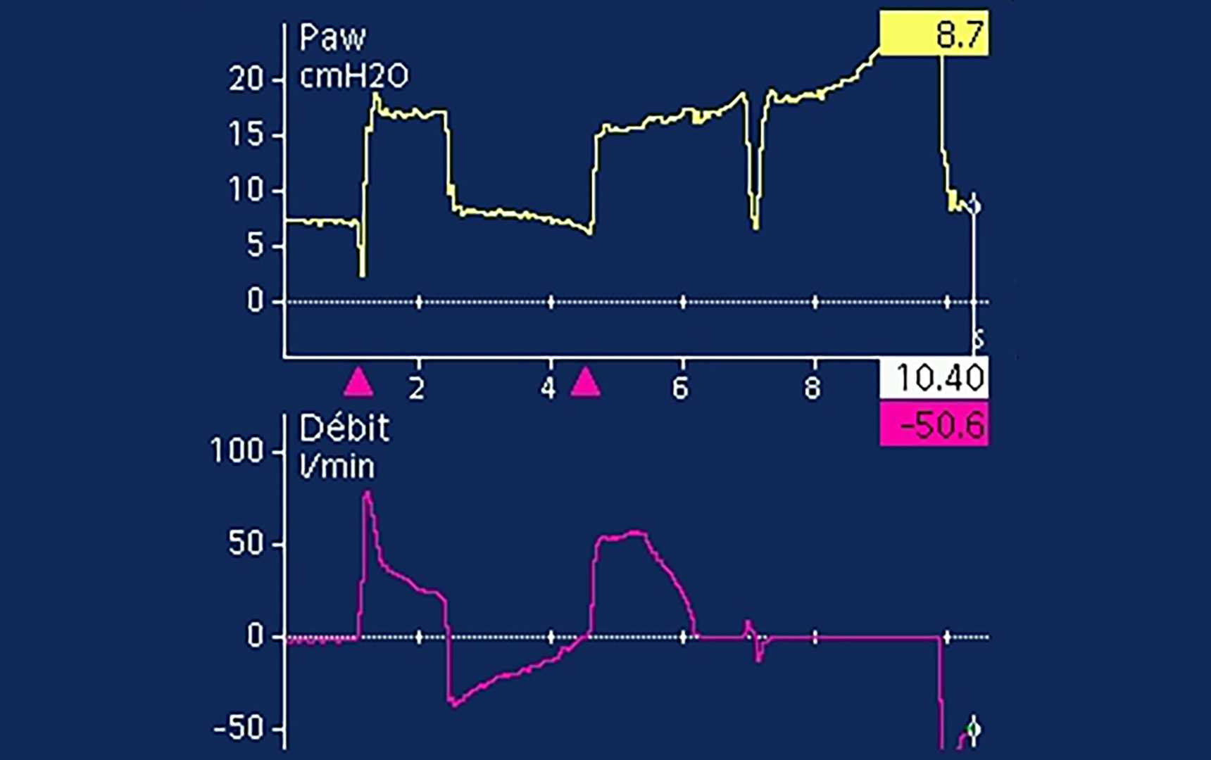
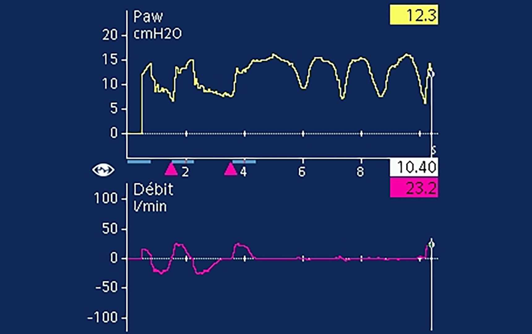
明白なプラトーが視認できる場合でも、呼気筋の活動が存在することは少なくありません(Soundoulounaki S, Akoumianaki E, Kondili E, et al. Airway pressure morphology and respiratory muscle activity during end-inspiratory occlusions in pressure support ventilation. Crit Care. 2020;24(1):467. Published 2020 Jul 28. doi:10.1186/s13054-020-03169-x6)。つまり、補助換気では∆PSTATは過大評価される可能性があります。したがって、低い∆PSTATは安全とみなすことができます。

Driving Pressure Is Associated with Outcome during Assisted Ventilation in Acute Respiratory Distress Syndrome.

Bellani G, Grassi A, Sosio S, et al. Driving Pressure Is Associated with Outcome during Assisted Ventilation in Acute Respiratory Distress Syndrome. Anesthesiology. 2019;131(3):594-604. doi:10.1097/ALN.0000000000002846

WHAT WE ALREADY KNOW ABOUT THIS TOPIC Higher driving pressure during controlled mechanical ventilation is known to be associated with increased mortality in patients with acute respiratory distress syndrome.Whereas patients with acute respiratory distress syndrome are initially managed with controlled mechanical ventilation, as they improve, they are transitioned to assisted ventilation. Whether higher driving pressure assessed during pressure support (assisted) ventilation can be reliably assessed and whether higher driving pressure is associated with worse outcomes in patients with acute respiratory distress syndrome has not been well studied. WHAT THIS ARTICLE TELLS US THAT IS NEW This study shows that in the majority of adult patients with acute respiratory distress syndrome, both driving pressure and respiratory system compliance can be reliably measured during pressure support (assisted) ventilation.Higher driving pressure measured during pressure support (assisted) ventilation significantly associates with increased intensive care unit mortality, whereas peak inspiratory pressure does not.Lower respiratory system compliance also significantly associates with increased intensive care unit mortality. BACKGROUND Driving pressure, the difference between plateau pressure and positive end-expiratory pressure (PEEP), is closely associated with increased mortality in patients with acute respiratory distress syndrome (ARDS). Although this relationship has been demonstrated during controlled mechanical ventilation, plateau pressure is often not measured during spontaneous breathing because of concerns about validity. The objective of the present study is to verify whether driving pressure and respiratory system compliance are independently associated with increased mortality during assisted ventilation (i.e., pressure support ventilation). METHODS This is a retrospective cohort study conducted on 154 patients with ARDS in whom plateau pressure during the first three days of assisted ventilation was available. Associations between driving pressure, respiratory system compliance, and survival were assessed by univariable and multivariable analysis. In patients who underwent a computed tomography scan (n = 23) during the stage of assisted ventilation, the quantity of aerated lung was compared with respiratory system compliance measured on the same date. RESULTS In contrast to controlled mechanical ventilation, plateau pressure during assisted ventilation was higher than the sum of PEEP and pressure support (peak pressure). Driving pressure was higher (11 [9-14] vs. 10 [8-11] cm H2O; P = 0.004); compliance was lower (40 [30-50] vs. 51 [42-61] ml · cm H2O; P < 0.001); and peak pressure was similar, in nonsurvivors versus survivors. Lower respiratory system compliance (odds ratio, 0.92 [0.88-0.96]) and higher driving pressure (odds ratio, 1.34 [1.12-1.61]) were each independently associated with increased risk of death. Respiratory system compliance was correlated with the aerated lung volume (n = 23, r = 0.69, P < 0.0001). CONCLUSIONS In patients with ARDS, plateau pressure, driving pressure, and respiratory system compliance can be measured during assisted ventilation, and both higher driving pressure and lower compliance are associated with increased mortality.

Increased Driving Pressure During Assisted Ventilation for Hypoxemic Respiratory Failure Is Associated with Lower ICU Survival: The ICEBERG Study.

Grassi A, Bianchi I, Teggia Droghi M, et al. Increased Driving Pressure During Assisted Ventilation for Hypoxemic Respiratory Failure Is Associated with Lower ICU Survival: The ICEBERG Study. Am J Respir Crit Care Med. Published online June 20, 2025. doi:10.1164/rccm.202411-2146OC

RATIONALE Driving pressure is marker of severity and a possible target for lung protection during controlled ventilation, but its value during assisted ventilation is unknown. Inspiratory holds provide an estimate of driving pressure (quasi-static). Expiratory holds provide an estimate of the inspiratory effort, useful to estimate the transpulmonary dynamic driving pressure. OBJECTIVES To assess the correlation between driving pressures measured during assisted ventilation and ICU outcomes. METHODS Multicenter prospective observational study. Patients with acute hypoxemic respiratory failure were enrolled within 48 hours of triggering the ventilator. Respiratory mechanics were measured daily and the variables of interest averaged over the first three days of partial assistance. ICU outcomes were collected until day 90. MEASUREMENTS AND MAIN RESULTS Two-hundred ninety-eight patients from 16 centers were enrolled. Tidal volume, peak airway pressure, positive-end-expiratory-pressure and inspiratory effort during the first three days of assisted ventilation did not differ between survivors and non-survivors. Quasi-static driving pressure and transpulmonary dynamic driving pressure were higher in non-survivors than in survivors (13 [11,14] vs 11 [9,13] cmH2O, p<0.001 and 19 [16,23] vs 16 [13,18] cmH2O, p<0.001, respectively), while compliance normalized to predicted body weight was lower (0.65 [0.54,0.84] vs 0.79 [0.64,0.97] ml/cmH2O/kg, p<0.001). Multivariable analysis confirmed the association with outcome. Over study days, static driving pressure significantly diverged between survivors and non-survivors. CONCLUSIONS During assisted ventilation driving pressure and normalized compliance are associated with ICU outcome, despite some overlap. Albeit our study does not allow to estimate if driving pressure is a marker of severity, or a cause of lung injury, it highlights the potential value of monitoring and targeting it during spontaneous assisted breathing.

End-inspiratory airway occlusion: a method to assess the pressure developed by inspiratory muscles in patients with acute lung injury undergoing pressure support.

Foti G, Cereda M, Banfi G, Pelosi P, Fumagalli R, Pesenti A. End-inspiratory airway occlusion: a method to assess the pressure developed by inspiratory muscles in patients with acute lung injury undergoing pressure support. Am J Respir Crit Care Med. 1997;156(4 Pt 1):1210-1216. doi:10.1164/ajrccm.156.4.96-02031

We evaluated the end-inspiratory occlusion maneuver as a means to estimate the inspiratory effort during pressure support ventilation (PS). In nine nonobstructed acute lung injury (ALI) patients, we applied four levels of PS (0, 5, 10, 15 cm H2O) to modify the inspiratory effort. End inspiratory occlusions (2 to 3 s) were performed at the end of each experimental period by pushing the inspiratory hold button of the ventilator (Servo 900 C; Siemens, Berlin, Germany). We took the difference between the end-inspiratory occlusion plateau pressure and the airway pressure before the occlusion (PEEP + PS) as an estimate of the inspiratory effort and called it PMI (Pmusc,index). From the esophageal pressure tracing we obtained a reference measurement of the pressure developed by the inspiratory muscles at end inspiration (Pmusc,ei) and of the pressure-time product per breath (PTP/b) and per minute (PTP/min). In each patient, PMI was correlated with Pmusc,ei (p < 0.01) and PTP/b (p < 0.01). A PMI threshold of 6 cm H2O detected PTP/min < 125 cm H2O s/min with a sensitivity of 0.89 and a specificity of 0.89. We conclude that PMI is a good estimate of the pressure developed by the inspiratory muscles in ALI patients and may be used to titrate PS level. The major advantage of PMI is that it can be obtained from the ventilator display without any additional equipment.

Plateau and driving pressure in the presence of spontaneous breathing.

Bellani G, Grassi A, Sosio S, Foti G. Plateau and driving pressure in the presence of spontaneous breathing. Intensive Care Med. 2019;45(1):97-98. doi:10.1007/s00134-018-5311-9

Reliability of plateau pressure during patient-triggered assisted ventilation. Analysis of a multicentre database.

Bianchi I, Grassi A, Pham T, et al. Reliability of plateau pressure during patient-triggered assisted ventilation. Analysis of a multicentre database. J Crit Care. 2022;68:96-103. doi:10.1016/j.jcrc.2021.12.002

PURPOSE An inspiratory hold during patient-triggered assisted ventilation potentially allows to measure driving pressure and inspiratory effort. However, muscular activity can make this measurement unreliable. We aim to define the criteria for inspiratory holds reliability during patient-triggered breaths. MATERIAL AND METHODS Flow, airway and esophageal pressure recordings during patient-triggered breaths from a multicentre observational study (BEARDS, NCT03447288) were evaluated by six independent raters, to determine plateau pressure readability. Features of "readable" and "unreadable" holds were compared. Muscle pressure estimate from the hold was validated against other measures of inspiratory effort. RESULTS Ninety-two percent of the recordings were consistently judged as readable or unreadable by at least four raters. Plateau measurement showed a high consistency among raters. A short time from airway peak to plateau pressure and a stable and longer plateau characterized readable holds. Unreadable plateaus were associated with higher indexes of inspiratory effort. Muscular pressure computed from the hold showed a strong correlation with independent indexes of inspiratory effort. CONCLUSION The definition of objective parameters of plateau reliability during assisted-breath provides the clinician with a tool to target a safer assisted-ventilation and to detect the presence of high inspiratory effort.

Airway pressure morphology and respiratory muscle activity during end-inspiratory occlusions in pressure support ventilation.

Soundoulounaki S, Akoumianaki E, Kondili E, et al. Airway pressure morphology and respiratory muscle activity during end-inspiratory occlusions in pressure support ventilation. Crit Care. 2020;24(1):467. Published 2020 Jul 28. doi:10.1186/s13054-020-03169-x

BACKGROUND The driving pressure of the respiratory system is a valuable indicator of global lung stress during passive mechanical ventilation. Monitoring lung stress in assisted ventilation is indispensable, but achieving passive conditions in spontaneously breathing patients to measure driving pressure is challenging. The accuracy of the morphology of airway pressure (Paw) during end-inspiratory occlusion to assure passive conditions during pressure support ventilation has not been examined. METHODS Retrospective analysis of end-inspiratory occlusions obtained from critically ill patients during pressure support ventilation. Flow, airway, esophageal, gastric, and transdiaphragmatic pressures were analyzed. The rise of gastric pressure during occlusion with a constant/decreasing transdiaphragmatic pressure was used to identify and quantify the expiratory muscle activity. The Paw during occlusion was classified in three patterns, based on the differences at three pre-defined points after occlusion (0.3, 1, and 2 s): a "passive-like" decrease followed by plateau, a pattern with "clear plateau," and an "irregular rise" pattern, which included all cases of late or continuous increase, with or without plateau. RESULTS Data from 40 patients and 227 occlusions were analyzed. Expiratory muscle activity during occlusion was identified in 79% of occlusions, and at all levels of assist. After classifying occlusions according to Paw pattern, expiratory muscle activity was identified in 52%, 67%, and 100% of cases of Paw of passive-like, clear plateau, or irregular rise pattern, respectively. The driving pressure was evaluated in the 133 occlusions having a passive-like or clear plateau pattern in Paw. An increase in gastric pressure was present in 46%, 62%, and 64% of cases at 0.3, 1, and 2 s, respectively, and it was greater than 2 cmH2O, in 10%, 20%, and 15% of cases at 0.3, 1, and 2 s, respectively. CONCLUSIONS The pattern of Paw during an end-inspiratory occlusion in pressure support cannot assure the absence of expiratory muscle activity and accurate measurement of driving pressure. Yet, because driving pressure can only be overestimated due to expiratory muscle contraction, in everyday practice, a low driving pressure indicates an absence of global lung over-stretch. A measurement of high driving pressure should prompt further diagnostic workup, such as a measurement of esophageal pressure.

Related articles. Get a deeper look