Voltar

Um guia compreensivo sobre ventilação não invasiva (VNI)

Artigo

Autor: Karjaghli Munir, Respiratory Therapist, Hamilton Medical Clinical Application Specialist

Data da primeira publicação: 01.03.2024

Descubra tudo o que precisa saber no nosso guia sobre ventilação VNI: desde a abreviatura médica até o que é ventilação não invasiva, quando utilizá-la, como selecionar a interface e as configurações, e muito mais.

Um guia compreensivo sobre ventilação não invasiva (VNI)

Terminologia, símbolos e abreviações

A ventilação não invasiva é comumente referida como VNI.

O termo ventilação não invasiva de pressão positiva (abreviado por NPPV ou NIPPV) foi previamente usado para distingui-la da ventilação não invasiva de pressão negativa, mas dada a raridade desta última atualmente, o termo mais simples VNI é mais conveniente. Uma vez que atualmente existe uma variedade de respiradores disponíveis para VNI, o uso do nome do produto BiPAP (Pressão positiva nas vias aéreas em dois níveis) como termo genérico para VNI deve ser evitado.

Outras abreviações são explicadas diretamente no texto.

Introdução

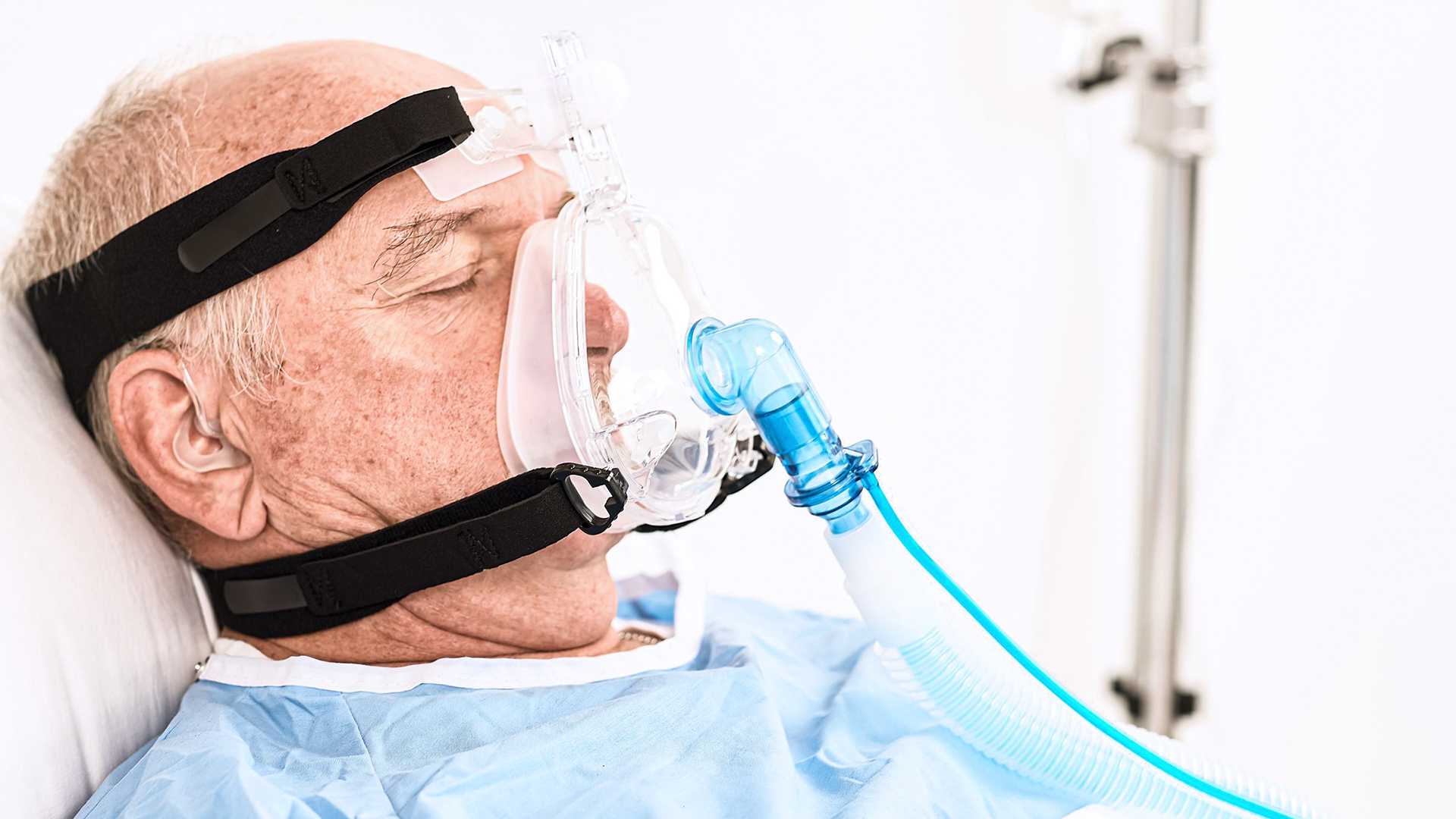
A ventilação não invasiva de pressão positiva envolve o fornecimento de oxigênio para os pulmões através de pressão positiva sem necessidade de intubação endotraqueal. Esta é usada tanto na insuficiência respiratória crônica quanto na aguda, mas exige a monitorização e titulação cuidadosas, de modo a garantir que é bem-sucedida e a evitar complicações (Rochwerg B, Brochard L, Elliott MW, et al. Official ERS/ATS clinical practice guidelines: noninvasive ventilation for acute respiratory failure. Eur Respir J. 2017;50(2):1602426. Published 2017 Aug 31. doi:10.1183/13993003.02426-20161).

Ao longo do último século, a ventilação não invasiva (VNI) teve grandes melhorias e tem sido usada para tratar a insuficiência respiratória de várias etiologias. Provou-se que ela é mais eficaz na prevenção da intubação em comparação com tratamento de oxigênio padrão no contexto agudo (Gong Y, Sankari A. Noninvasive Ventilation. In: StatPearls. Treasure Island (FL): StatPearls Publishing; December 11, 2022.2​).

O suporte respiratório pode ser fornecido usando dispositivos de pressão positiva contínua nas vias aéreas (CPAP) ou aqueles que fornecem pressão positiva nas vias aéreas em dois níveis (ventilação com suporte de pressão, PSV). Para fins deste artigo, o nome VNI abrange tanto CPAP quanto PSV (Gong Y, Sankari A. Noninvasive Ventilation. In: StatPearls. Treasure Island (FL): StatPearls Publishing; December 11, 2022.2​).

Paciente adulto com máscara VNI Pulmodyne
Ilustração para o capítulo “Quais são os benefícios de VNI?”

Quais são os benefícios? Objetivos e benefícios em usar a VNI

Podemos dividir os objetivos e benefícios da VNI no contexto de cuidados a longo prazo e agudos, da seguinte forma.

Benefícios da VNI em ambientes de cuidados agudos (Egan's fundamentals of respiratory care e-book3):

  • Melhorar a troca gasosa
  • Evitar a intubação
  • Diminuir a mortalidade
  • Diminuir o período de tempo no respirador
  • Diminuir a duração da hospitalização
  • Diminuir a incidência de pneumonia associada à ventilação
  • Aliviar os sintomas da angústia respiratória
  • Melhorar a sincronização do paciente com o respirador
  • Maximizar o conforto do paciente


Benefícios da VNI em ambientes de cuidados a longo prazo (Egan's fundamentals of respiratory care e-book3​):

  • Aliviar ou melhorar os sintomas
  • Melhorar a qualidade de vida
  • Evitar a hospitalização
  • Aumentar a sobrevivência
  • Melhorar a mobilidade
Ilustração para o capítulo “Como funciona a VNI?”

Como funciona o VNI?

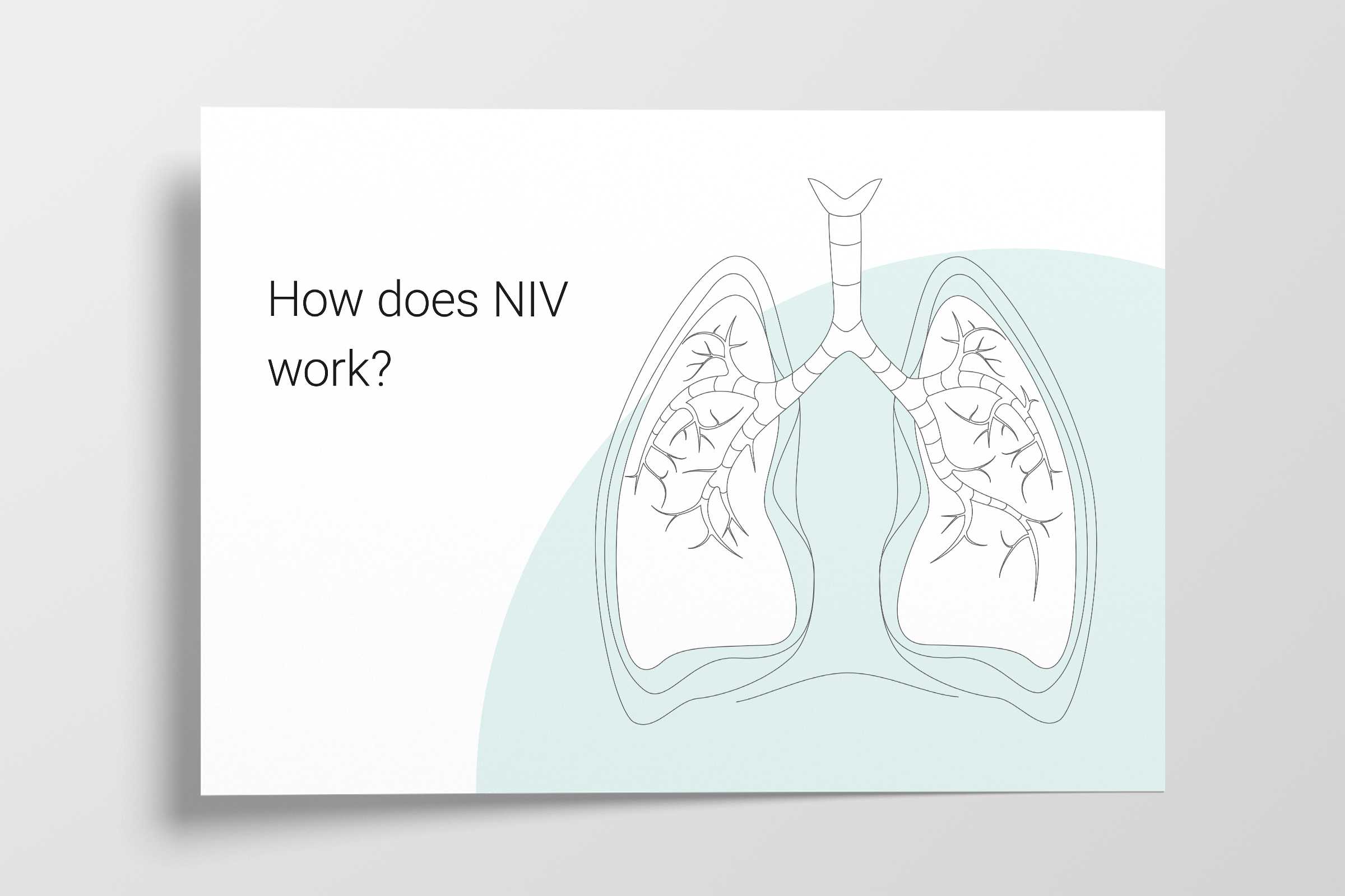
O funcionamento da ventilação não invasiva consiste em criar pressão positiva nas vias aéreas, ou seja, a pressão fora dos pulmões é maior do que a pressão dentro dos pulmões. Isto faz com que o ar seja forçado a entrar nos pulmões (pelo gradiente de pressão), diminuindo o esforço respiratório e reduzindo o trabalho respiratório.

Ela também ajuda a manter o tórax e os pulmões expandidos, aumentando a capacidade residual funcional (a quantidade de ar que permanece nos pulmões após a expiração) após a expiração (corrente) normal; este é o ar nos alvéolos disponível para a troca gasosa (Rochwerg B, Einav S, Chaudhuri D, et al. The role for high flow nasal cannula as a respiratory support strategy in adults: a clinical practice guideline. Intensive Care Med. 2020;46(12):2226-2237. doi:10.1007/s00134-020-06312-y4).

Paciente adulto com máscara VNI Pulmodyne

Modos VNI

Os modos seguintes são não invasivos: 

  • CPAP: Significa pressão positiva contínua nas vias aéreas
  • VNI (PSV): Também designado por modo CPAP em dois níveis ou modo de pressão em dois níveis
  • VNI-ST (espontânea/temporizada S/T): Significa ventilação não invasiva espontânea/temporizada

CPAP

CPAP tem como objetivo fornecer um nível contínuo de pressão positiva ao longo das fases inspiratória e expiratória de respiração.

Melhora a oxigenação, abrindo as vias aéreas colapsadas, melhorando a capacidade residual funcional (CRF) e melhorando a pré-carga e pós-carga no edema pulmonar cardiogênico (Bello G, De Santis P, Antonelli M. Non-invasive ventilation in cardiogenic pulmonary edema. Ann Transl Med. 2018;6(18):355. doi:10.21037/atm.2018.04.395​).

CPAP melhora a complacência pulmonar e, portanto, reduz o esforço necessário para respirar, evitando o colapso alveolar e neutralizando a PEEP intrínseca excessiva observada em condições obstrutivas dos pulmões, como a DPOC (Bello G, De Santis P, Antonelli M. Non-invasive ventilation in cardiogenic pulmonary edema. Ann Transl Med. 2018;6(18):355. doi:10.21037/atm.2018.04.395​, Davidson AC, Banham S, Elliott M, et al. BTS/ICS guideline for the ventilatory management of acute hypercapnic respiratory failure in adults [published correction appears in Thorax. 2017 Jun;72 (6):588]. Thorax. 2016;71 Suppl 2:ii1-ii35. doi:10.1136/thoraxjnl-2015-2082096).

A ter em conta: Se ΔPsuporte / ∆Pinsp for zero em VNI e VNI-ST, o respirador funcionará como um sistema comum de CPAP.

Ilustração de CPAP e VNI
Adaptado de Hess, Dean R. e Robert M. Kacmarek. Essentials of mechanical ventilation. McGraw Hill Education, 2019.
Ilustração de CPAP e VNI
Adaptado de Hess, Dean R. e Robert M. Kacmarek. Essentials of mechanical ventilation. McGraw Hill Education, 2019.

VNI (PSV)

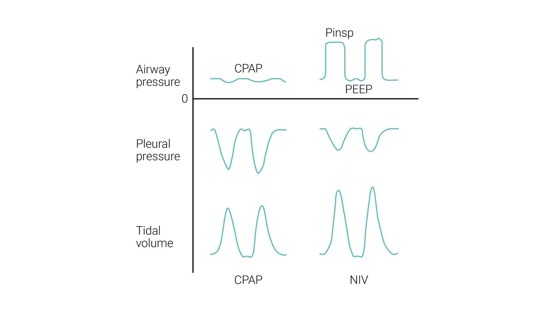
VNI (PSV) tem como objetivo fornecer dois níveis de suporte de pressão positiva nas vias aéreas. O nível menor é semelhante a CPAP; no entanto, é mais frequentemente designada por pressão positiva no final da expiração nas vias aéreas (PEEP), pois está presente apenas na fase expiratória da respiração. 

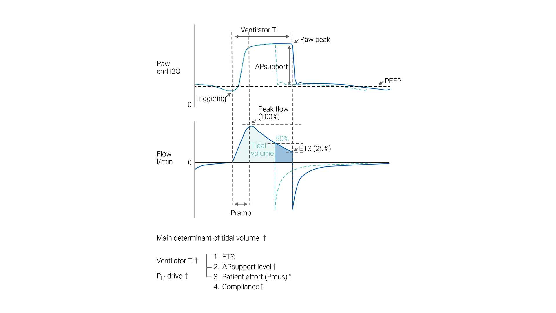
O esforço inspiratório do paciente é assistido pelo respirador em um nível predefinido de pressão inspiratória (ΔPsuporte). A inspiração é iniciada e alternada pelo esforço do paciente. Durante a VNI, o paciente determina a frequência respiratória, o tempo inspiratório e o volume corrente.

O tamanho do ciclo respiratório (volume corrente) gerado em um determinado paciente depende da configuração de ΔPsuporte — quanto maior for a configuração de ΔPsuporte, maior o volume corrente.

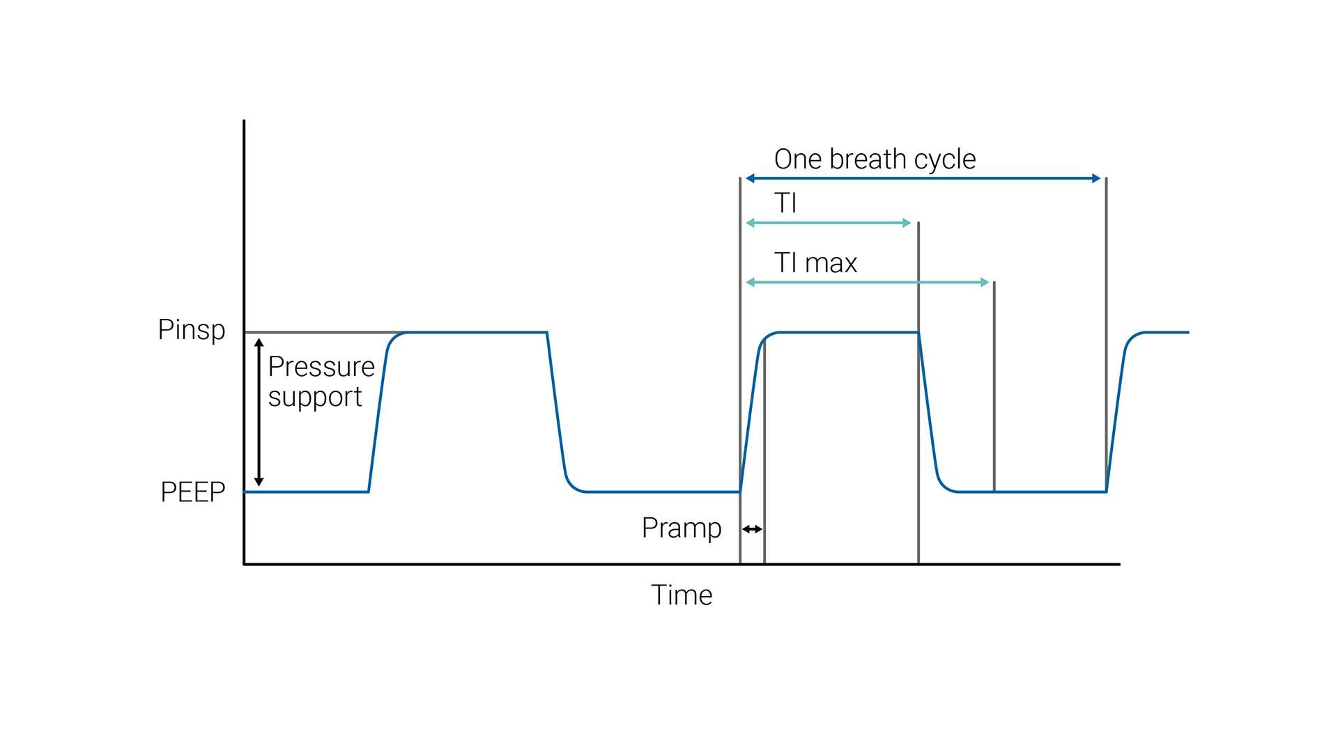
A ETS (sensibilidade de disparo expiratório) determina o tempo inspiratório espontâneo, alternando a expiração assim que o fluxo inspiratório diminuir para uma porcentagem pré-ajustada do pico de fluxo inspiratório.

Caso os critérios de ETS não sejam cumpridos (fuga), o tempo inspiratório também pode ser limitado por Ti máx (tempo inspiratório máximo). 

Ilustração da pressão e fluxo
Adaptado de Bellani, Giacomo. Mechanical ventilation from pathophysiology to clinical evidence. Cham, Switzerland: Springer, 2022.
Ilustração da pressão e fluxo
Adaptado de Bellani, Giacomo. Mechanical ventilation from pathophysiology to clinical evidence. Cham, Switzerland: Springer, 2022.

VNI-ST

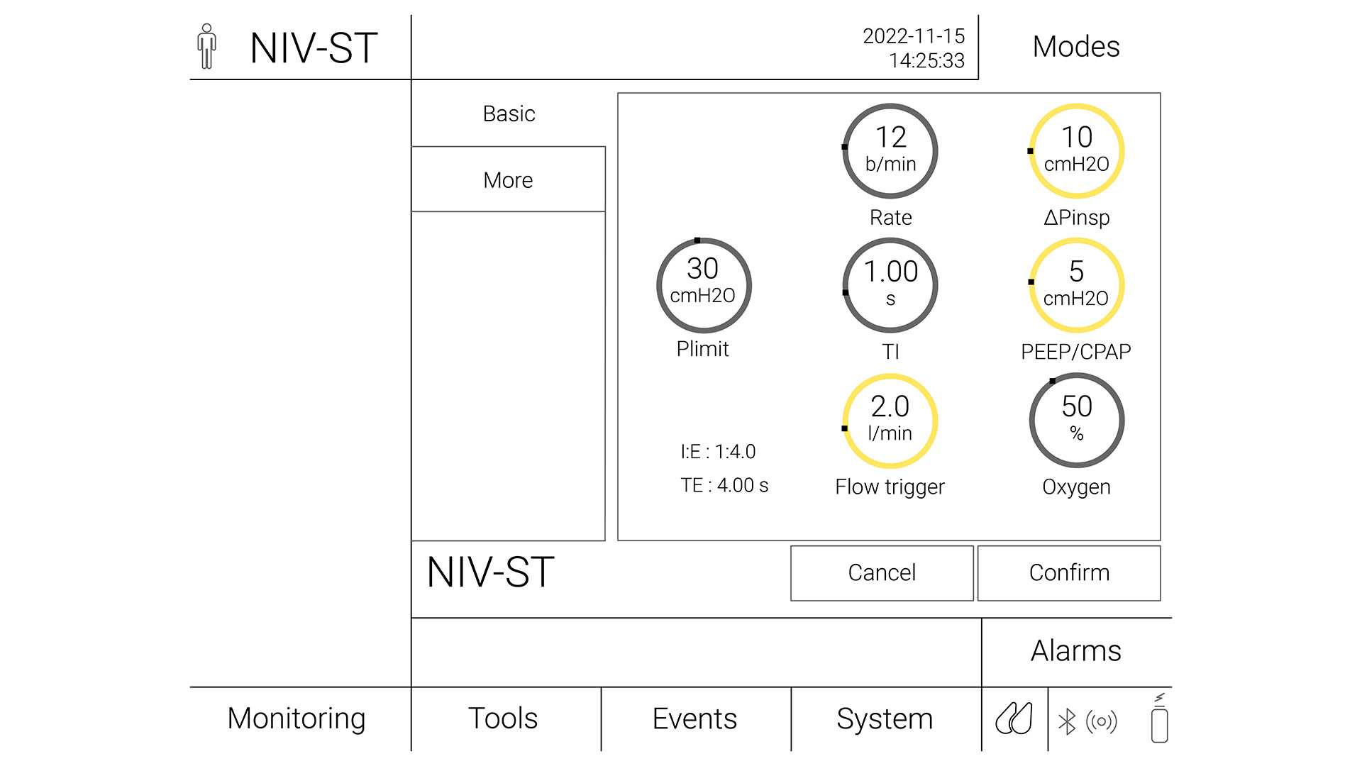
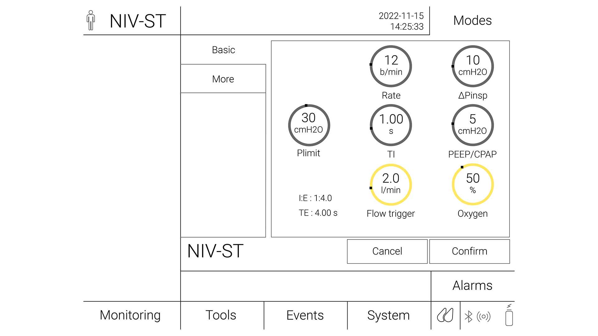
No modo S/T, o clínico configura a pressão inspiratória (∆Pinsp) e a pressão expiratória (PEEP), a frequência respiratória e o tempo inspiratório. O paciente pode iniciar ciclos respiratórios que são suportados até ao nível ∆Pinsp, como no modo VNI, mas se o paciente não fizer um esforço inspiratório em um intervalo definido (que é definido pela frequência respiratória definida), a máquina inicia a inspiração até ao nível ∆Pinsp definido. ∆Pinsp alterna, de seguida, para PEEP com base no período de tempo inspiratório.

A VNI-ST com a frequência de suporte é útil em caso de apneia ou ciclo respiratório periódico (Teaching Pearls in Noninvasive Mechanical Ventilation: Key Practical Insights7).

Ilustração de VNI-ST

Modo de suporte de pressão vs. CPAP/BPAP

Os modos VNI e VNI-ST são tipos de ventilação não invasiva de pressão positiva. O suporte de pressão é geralmente considerado como sendo o mesmo que CPAP/BiPAP, exceto o fato de o suporte de pressão ser fornecido por um respirador e BiPAP ser fornecido por um respirador não invasivo.

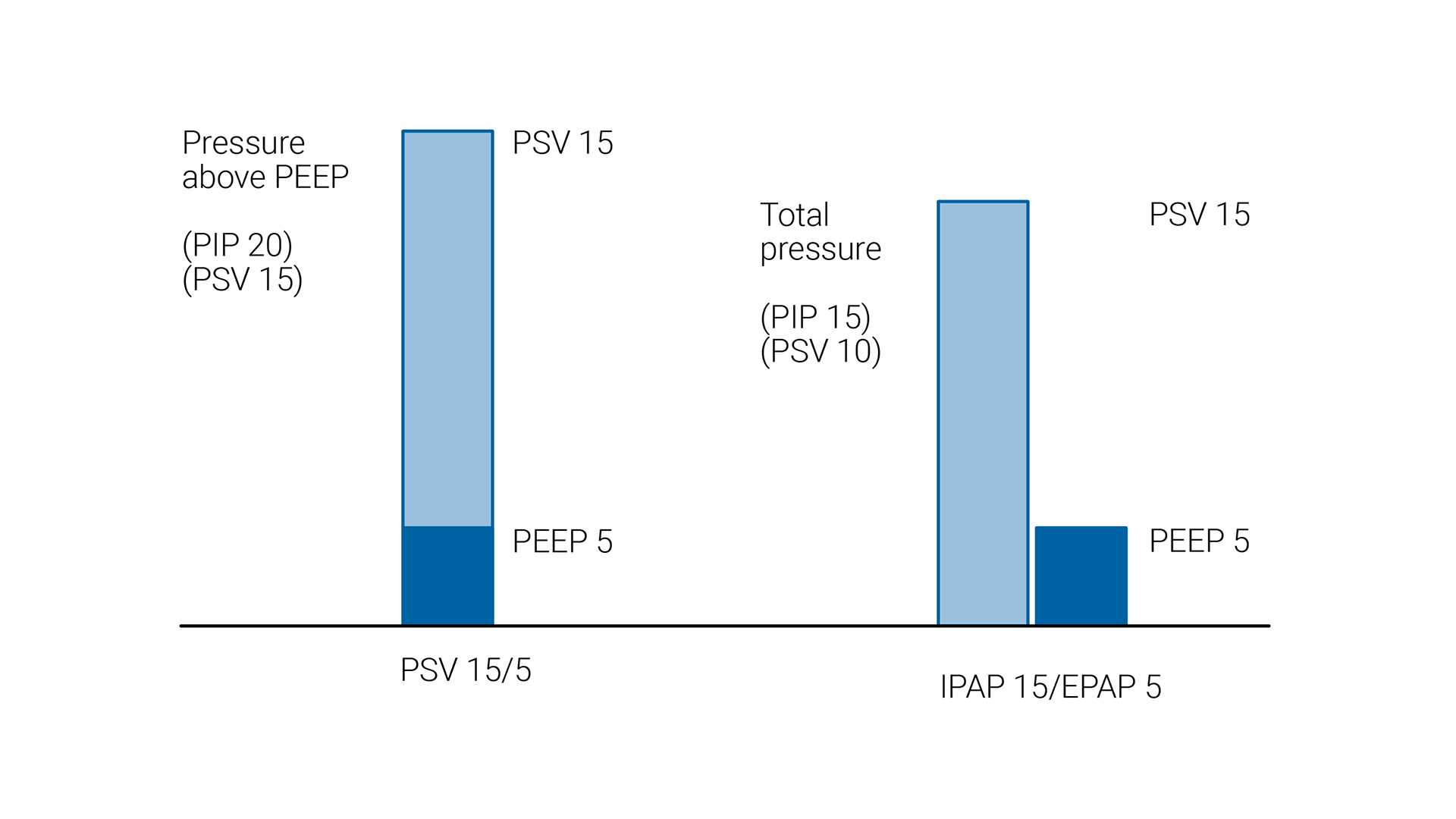
Em VNI e VNI-ST, o nível de suporte de pressão é aplicado como pressão acima da PEEP de linha basal. No entanto, a abordagem é diferente com respiradores de dois níveis, em que a PIPVA (pressão positiva inspiratória nas vias aéreas) e a PEPVA (pressão positiva expiratória nas vias aéreas) são definidas. Nesta configuração, a diferença entre PIPVA e PEPVA é o nível de suporte de pressão.

Comparação da PSV e PIPVA
Comparação da ventilação com suporte de pressão (PSV), como nos respiradores de terapia intensiva, e da pressão inspiratória positiva nas vias aéreas (PIPVA) com um respirador de dois níveis. Note que a PIPVA é a pressão inspiratória de pico (PIP) e inclui a pressão expiratória positiva nas vias aéreas (PEPVA), enquanto na PSV o suporte de pressão é fornecido além da pressão expiratória positiva ao final da expiração (PEEP).

Quando considerar a ventilação não invasiva?

De modo a minimizar o risco de falhas ou complicações, cada paciente deve ser avaliado corretamente quanto à sua adequação para receber a VNI com segurança.

As indicações para o uso da VNI incluem os pacientes com:

  • Dispneia
  • Taquicardia
  • Uso dos músculos respiratórios acessórios
  • Respiração “pela barriga” ou respiração abdominal paradoxal
  • PaCO2 > 45 mmHg e pH < 7,35 (Ozyilmaz E, Ugurlu AO, Nava S. Timing of noninvasive ventilation failure: causes, risk factors, and potential remedies. BMC Pulm Med. 2014;14:19. Published 2014 Feb 13. doi:10.1186/1471-2466-14-198​,Scala R, Pisani L. Noninvasive ventilation in acute respiratory failure: which recipe for success?. Eur Respir Rev. 2018;27(149):180029. Published 2018 Jul 11. doi:10.1183/16000617.0029-20189)

Os pacientes devem ser monitorados cuidadosamente durante as primeiras 24 horas após iniciarem a VNI, uma vez que este é o período com a maior taxa de insucesso do tratamento. Embora os pontos de dados na apresentação, como uma FR (frequência respiratória) alta, valores baixos de pH arterial ou PaO2/FiO2 baixa, possam ajudar a prever a falha, o indicador mais forte do insucesso do tratamento durante este período é a não demonstração de um melhoramento nestes parâmetros, 1–2 horas após o início do tratamento de VNI (Scala R, Pisani L. Noninvasive ventilation in acute respiratory failure: which recipe for success?. Eur Respir Rev. 2018;27(149):180029. Published 2018 Jul 11. doi:10.1183/16000617.0029-20189).

Indicações e recomendações para a NPPV

Clinical indication

Certainty of evidence

Recommendation

Hypercapnia with COPD exacerbation

High

Strong recommendation for

Cardiogenic pulmonary edema (CPE)

Moderate

Strong recommendation for

Immunocompromised

Moderate

Conditional recommendation for

Post-operative patients

Moderate

Conditional recommendation for

Palliative care

Moderate

Conditional recommendation for

Trauma

Moderate

Conditional recommendation for

Weaning in hypercapnic patients

Moderate

Conditional recommendation for

Post-extubation respiratory failure

Low

Conditional recommendation against

Obesity hypoventilation syndrome (OHS)

Low

Conditional recommendation for

Neuromuscular disease and chest wall disease

Low

Conditional recommendation for

Prevention of hypercapnia in COPD exacerbation

Low

Conditional recommendation against

Post-extubation in high-risk patients (prophylaxis)

Low

Conditional recommendation for

De novo respiratory failure

No certain evidence

No recommendation made

Acute asthma exacerbation

No certain evidence

No recommendation made

Contraindicações à ventilação não invasiva

Absolutas:

  • A necessidade de intubação emergente (ou seja, paragem respiratória ou cardíaca, angústia respiratória grave, arritmia cardíaca instável)

Relativas:

  • A falência de órgãos não respiratórios potencialmente fatal
  • Encefalopatia grave (ou seja, escala de coma de Glasgow < 10)
  • Sangramento gastrointestinal superior grave
  • Instabilidade hemodinâmica
  • Cirurgia neurológica ou facial, trauma ou deformidade
  • Obstrução significativa das vias aéreas (ou seja, massa laríngea ou tumor traqueal)
  • Incapacidade em cooperar, proteger as vias aéreas ou limpar secreções (ou seja, pacientes em alto risco de aspiração)
  • Duração prolongada antecipada da ventilação mecânica (ou seja, ≥ 4 a 7 dias)
  • Anastomose gástrica ou esofágica recente (ECG: A escala de coma de Glasgow e a distensão gástrica ou esofágica por ar podem aumentar o risco de deiscência anastomóticaA)
  • Várias contraindicações
  • Suporte insuficiente da equipe

(Evans TW. International Consensus Conferences in Intensive Care Medicine: non-invasive positive pressure ventilation in acute respiratory failure. Organised jointly by the American Thoracic Society, the European Respiratory Society, the European Society of Intensive Care Medicine, and the Société de Réanimation de Langue Française, and approved by the ATS Board of Directors, December 2000. Intensive Care Med. 2001;27(1):166-178. doi:10.1007/s00134000072110)

Ilustração para o capítulo “Qual é a relevância clínica de VNI?”

Qual é a relevância clínica de VNI? Efeitos fisiológicos da ventilação não invasiva

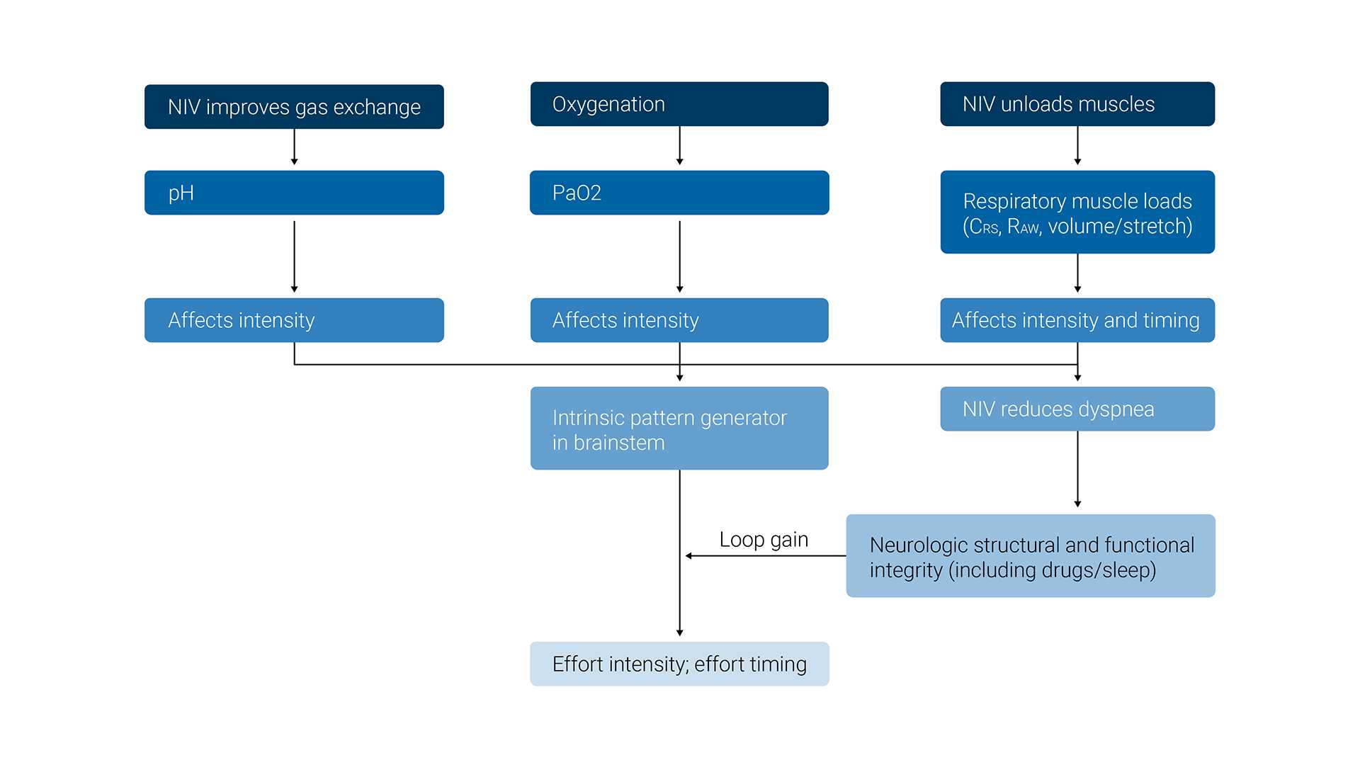
O principal efeito desejado da VNI é manter os níveis adequados de PO2 e PCO2 no sangue arterial e, ao mesmo tempo, aliviar a carga dos músculos inspiratórios.

Os efeitos fisiológicos da ventilação não invasiva são os seguintes: 

  • Aumenta a ventilação alveolar por minuto
  • Alivia a carga dos músculos respiratórios
  • Repõe o sistema de controle ventilatório
  • Melhora o recrutamento alveolar e da troca gasosa
  • Mantém a patência das vias aéreas
  • Reduz cargas de disparo de AutoPEEP
  • Reduz o risco de lesão pulmonar induzida pelo respirador

(MacIntyre NR. Physiologic Effects of Noninvasive Ventilation. Respir Care. 2019;64(6):617-628. doi:10.4187/respcare.0663511​)

Ilustração dos efeitos fisiológicos de VNI
Adaptado de MacIntyre, Neil R. Physiologic effects of noninvasive ventilation. Respiratory care 64.6 (2019): 617-628
Ilustração dos efeitos fisiológicos de VNI
Adaptado de MacIntyre, Neil R. Physiologic effects of noninvasive ventilation. Respiratory care 64.6 (2019): 617-628
Ilustração para o capítulo “Como iniciar um paciente na VNI?”

Como iniciar um paciente na VNI?

  • Formação: planeje um treinamento adequado para toda a equipe com um protocolo calibrado
  • Ambiente: escolha uma configuração adequada para iniciar a VNI de acordo com a severidade da IRA
  • Indicação: selecione os pacientes de acordo com a experiência da equipe, local, disponibilidade de intubação, status de não intubação e a probabilidade de sucesso
  • Informação: explique a técnica a pacientes competentes para melhorar a sua complacência
  • Equipamento: escolha a(s) interface(s) que melhor se adequam à anatomia facial; considere também alternar entre diferentes interfaces para melhorar o conforto. Escolha um respirador com uma boa compensação de fuga de ar que exiba curvas de fluxo/pressão/volume
  • Iniciar a ventilação: escolha um modo de pressão (ou seja, suporte de pressão) com PEEP. Inicie com pressões baixas e, de seguida, aumente gradualmente, dependendo do conforto. Configure os alarmes essenciais e FiO2 adequada. Aperte suficientemente as faixas da interface, de forma a evitar fugas, sem apertá-las demasiado
  • Monitorização da ventilação: verifique o status clínico, monitore a SpO2, meça a gasometria arterial periodicamente. Reponha o respirador de acordo com a sincronização do paciente com o respirador, conforto e fugas. Evite lesões na pele (ou seja, dispositivos de proteção, interfaces giratórias). Considere a umidificação. Considere cuidadosamente a sedação. Considere a redução de secreções, se necessário.

(Non-invasive ventilation and weaning: principles and practice12)

Condições para iniciar a VNI

  • Escolha um local com monitorização adequada, com base na severidade da condição do paciente. No mínimo, deve ser fornecida a oximetria de pulso contínua.
  • A otimização da posição do paciente também desempenha um papel fundamental para garantir o conforto durante a VNI (Non–Invasive Ventilation Guidelines for Adult Patients With Acute Respiratory Failure: A Clinical Practice Guideline.13). Sugere-se uma posição sentada ou semirrecostada durante a VNI, para garantir um alto nível de conforto aos pacientes. Uma posição deitada de lado pode ajudar a remover a pressão de um abdômen pendente, como no caso de gravidez ou obesidade (Non–Invasive Ventilation Guidelines for Adult Patients With Acute Respiratory Failure: A Clinical Practice Guideline.13​). Recentemente, o uso do posicionamento prono foi introduzido em pacientes com IRA, especialmente aqueles com COVID-19 (Ehrmann S, Li J, Ibarra-Estrada M, et al. Awake prone positioning for COVID-19 acute hypoxaemic respiratory failure: a randomised, controlled, multinational, open-label meta-trial. Lancet Respir Med. 2021;9(12):1387-1395. doi:10.1016/S2213-2600(21)00356-814). A análise deste tratamento de resgate é explicada de forma melhor no último parágrafo sobre a pandemia de COVID-19.
  • Selecione um respirador e uma interface com o tamanho adequado, e garanta que a interface é compatível com o tipo de respirador a ser usado.
Ilustração de um paciente na posição semirrecostada
Ilustração para o capítulo “Como escolher a interface de VNI correta?”

Como escolher a interface de VNI correta?

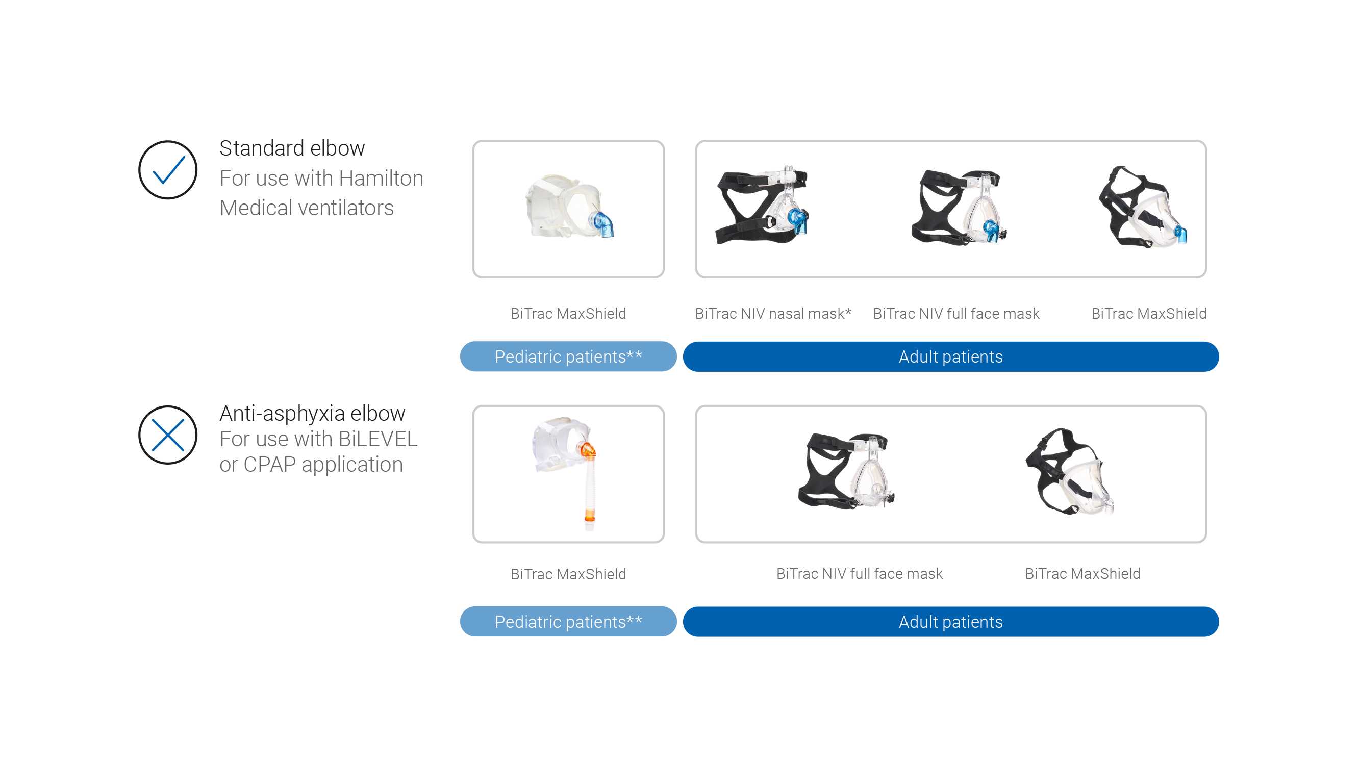
Selecione a máscara concebida para ser utilizada com um respirador de terapia intensiva (sem porta de fuga ou válvulas de arrasto). 

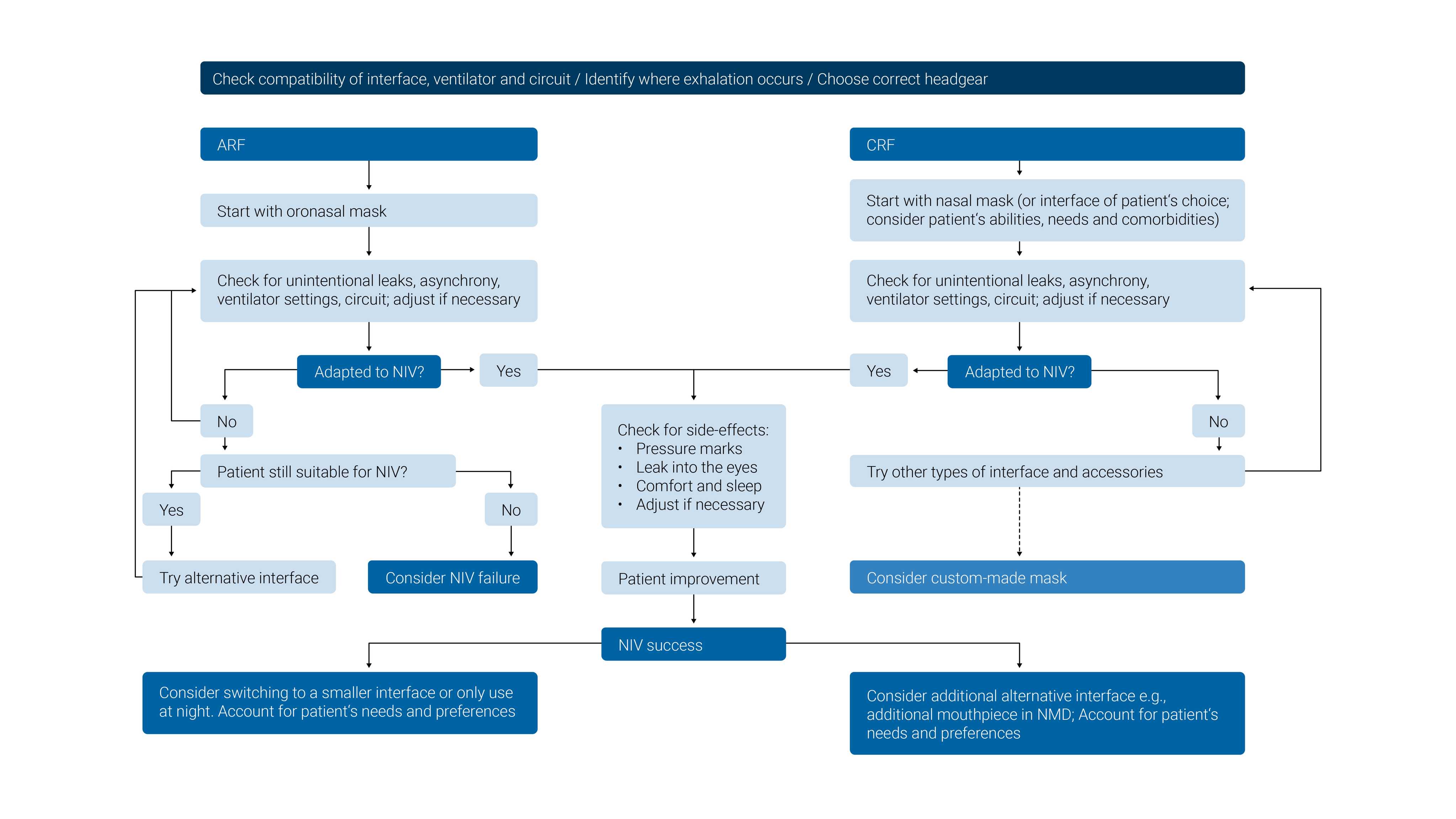
O tamanho errado da máscara, o posicionamento incorreto ou uma vedação insuficiente da máscara podem causar desconforto ao paciente e ferimentos por pressão. É importante escolher uma interface que tenha o tamanho adequado para cada paciente, avaliar o ajuste e o posicionamento da máscara no rosto e reposicionar a máscara conforme necessário para minimizar fugas. 

A ter em conta: As interfaces de VNI com válvulas de arrasto foram concebidas para respiradores não invasivos que usam um circuito de alça única. A válvula de arrasto é necessária para evitar a asfixia em caso de falha do respirador ou de desconexão do tubo. As máscaras com portas de fuga apenas devem ser usadas com respirador de circuito de alça única e não devem ser usadas com respiradores Hamilton Medical.

(Simonds, A. K. (Ed.). (2015). ERS practical handbook of noninvasive ventilation. European Respiratory Society.15)

A photo of the selection of NIV interfaces available at Hamilton Medical
A diagram tree of the compatibility of NIV interfaces
Adapted from Simonds, Anita K., ed. ERS practical handbook of noninvasive ventilation. European Respiratory Society, 2015.
A diagram tree of the compatibility of NIV interfaces
Adapted from Simonds, Anita K., ed. ERS practical handbook of noninvasive ventilation. European Respiratory Society, 2015.
Ilustração para o capítulo “Como configurar a VNI?”

Como configurar a VNI?

  • Ligue o respirador e umidificador e, de seguida, execute o teste de estanqueidade e calibração
  • Conecte a interface ao circuito
  • Explique o procedimento e a razão para o tratamento ao paciente e responda a quaisquer perguntas sobre a VNI antes de colocar a máscara no paciente
Ilustração de uma tela do respirador com o teste e a calibração para VNI-ST

Umidificação durante a VNI

Recomendações (Holanda MA, Reis RC, Winkeler GF, Fortaleza SC, Lima JW, Pereira ED. Influence of total face, facial and nasal masks on short-term adverse effects during noninvasive ventilation. J Bras Pneumol. 2009;35(2):164-173. doi:10.1590/s1806-3713200900020001016​, American Association for Respiratory Care, Restrepo RD, Walsh BK. Humidification during invasive and noninvasive mechanical ventilation: 2012. Respir Care. 2012;57(5):782-788. doi:10.4187/respcare.0176617​):

  • Todos os circuitos de VNI devem ser ativamente umidificados
  • Os HMEs (trocadores de calor e umidade) não são recomendados para a VNI
  • As temperaturas dos gases durante a VNI devem ser baseadas no conforto do paciente
  • O gás umidificado, aquecido a uma temperatura que seja confortável para o paciente (normalmente, a aprox. 30 °C), deve ser o padrão ao usar a VNI

Concordância e motivação do paciente

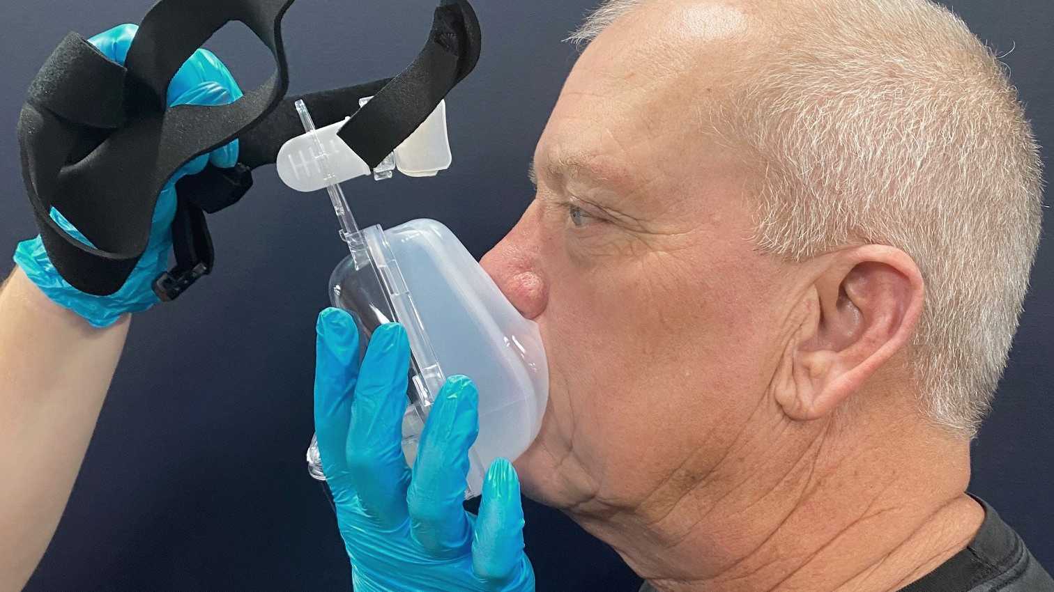
A dispneia pode causar sentimentos de ansiedade e medo. Por isso, o profissional de saúde ou o paciente deve segurar a máscara no lugar ao aplicá-la pela primeira vez.

Desta forma, a máscara pode ser removida rapidamente se o paciente começar a entrar em pânico ou precisar comunicar. A estratégia de iniciar com pressões baixas pode ajudar os pacientes a se adaptar mais rapidamente à VNI.

Paciente e máscara VNI
Ilustração para o capítulo “Como ajustar as configurações da VNI?”

Como ajustar as configurações? Configurações iniciais do respirador para VNI

  • As pressões de ventilação devem ser configuradas no valor mais baixo possível no início, especialmente se o paciente não estiver familiarizado com a sensação de ventilação de pressão positiva (PEEP 4–6 cmH2O, ∆Pinsp 6–10 cmH2O)
  • As pressões de ventilação podem então ser configuradas em pequenos incrementos durante 1 a 2 minutos, até o Vcorr exalado ser de 6 a 8 ml/kg de peso corporal previsto ou até a angústia respiratória melhorar
  • Ajuste a FiO2 para manter a SpO2 no intervalo desejado (88%–95%)
  • Disparo i-F: configure a sensibilidade de disparo o suficiente para permitir o disparo sem início espontâneo, mesmo na presença de fugas
  • Os picos de pressão das vias aéreas superiores a 30 cmH2O raramente são necessários e o ideal é evitá-los. As configurações de pressão superiores a 30 cmH2O podem forçar o ar pelo esôfago e para o estômago (O Limite P não deve exceder 30 cmH2O ou a PEEP 8 cmH2O sem a verificação por um especialistaC​)
  • Configure a frequência de suporte para 12–16 bpm (2 bpm abaixo da frequência respiratória em repouso)
  • Configure uma relação I:E e um tempo inspiratório adequados para a condição apresentada pelo paciente (Tinsp e Rel I:E (1:2–1:3 (DPOC) ou 1:1 (NMD/SOH)) configurados por um profissional competenteD​)
  • ETS: 40%–50%
  • Prampa (rampa de pressão): deve ser ajustada para uma inclinação mais rápida, evitando, ao mesmo tempo, o fluxo de pico excessivo
  • O tempo inspiratório máximo é configurado para 0,25 s acima do tempo inspiratório real; se você não tiver certeza, inicie em 1,5 s

(Developed from BTS/ICS guidelines (https://www.brit-thoracic.org.uk/document-library/clinical-information/acute-hypercapnic-respiratory-failure/bts-guidelines-for-ventilatory-management-of-ahrf/)B​)

Image of ventilator settings for NIV

Configuração dos alarmes para VNI

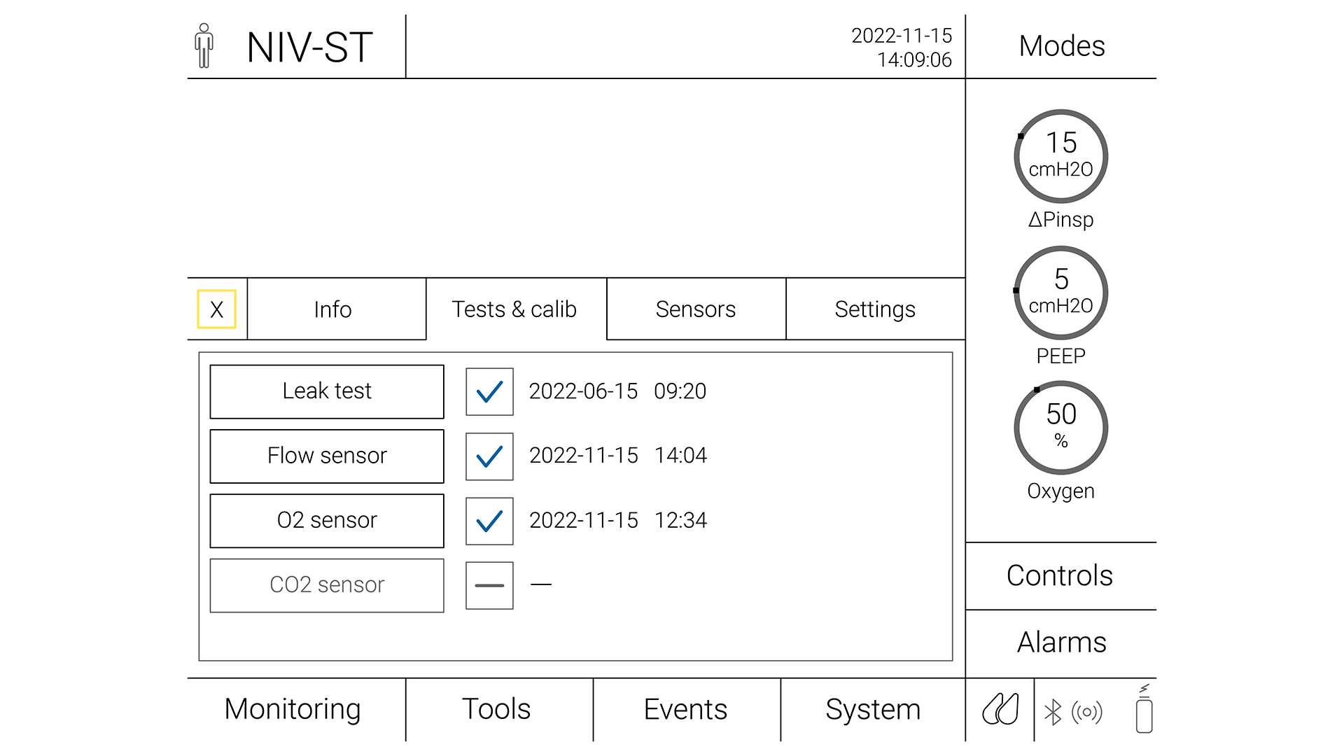
Os alarmes de emergência são importantes para reconhecer a deterioração na condição do paciente e alertar a equipe nas seguintes situações:

  • Apneia: quando o paciente para de respirar
  • Frequência respiratória alta: quando a frequência respiratória do paciente excede o valor ajustado
  • Frequência respiratória baixa: quando a frequência respiratória do paciente fica abaixo de um valor ajustado
  • Volume corrente alto: quando o volume corrente do paciente excede um valor alvo
  • Volume corrente baixo: quando o volume corrente do paciente fica abaixo de um valor alvo
Ilustração para o capítulo “Como devo monitorar os pacientes em VNI?”

Como devo monitorar os pacientes em VNI?

(Developed from BTS/ICS guidelines (https://www.brit-thoracic.org.uk/document-library/clinical-information/acute-hypercapnic-respiratory-failure/bts-guidelines-for-ventilatory-management-of-ahrf/)B​)

  • Reavalie frequentemente quanto à tolerância e eficácia da VNI (pelo menos, a cada 30 min) durante as primeiras 1 a 2 horas
  • Monitorização cardíaca e de SpO2 contínuas por, pelo menos, as primeiras 12 horas. Certifique-se de que os parâmetros PaCO2, PaO2 e SpO2 estão ajustados
  • Alterar as configurações da VNI: se a PaCO2 permanecer alta, aumente o volume corrente (Vcorr), aumentando a ∆Pinsp. Se o paciente continuar hipóxico, aumente a PEEP ou a FiO2 (lembre-se de que talvez seja necessário aumentar a ∆Pinsp para manter o Vcorr entre 6–8 ml/kg ou para os sinais de angústia respiratória melhorarem), atualize a prescrição de VNI e repita a ABG (gasometria arterial) uma hora após a alteração de qualquer configuração (Aumente a ∆Pinsp por 10–30 minutos. A Ppico não deve exceder 30 cmH2O ou a PEEP 8 cmH2O sem a verificação por um especialistaE​)
  • Verifique a tolerância do paciente com as configurações iniciais e avalie quanto à dispneia e assincronia
  • Monitore o paciente quanto ao risco de possível insucesso da VNI
  • Use os critérios pré-especificados para o agravamento
  • Não atrase uma intubação indicada

Falha de monitorização durante a ventilação não invasiva

O insucesso da VNI tem sido geralmente definido como a necessidade de intubação devido à inexistência de um melhoramento na gasometria arterial e nos parâmetros clínicos, ou morte (Teaching Pearls in Noninvasive Mechanical Ventilation: Key Practical Insights7​). É muito importante identificar os pacientes que correm o risco de não serem bem-sucedidos na VNI, pois um atraso inadequado na intubação pode causar um aumento na morbilidade e na mortalidade.

Preditores de sucesso:

  • Aumento na oxigenação arterial
  • Diminuição na frequência respiratória
  • Diminuição na dispneia
  • Aumento significativo na PaO2/FiO2 (Pressão parcial de oxigênio/Fração de oxigênio inspirado)

Preditores de insucesso:

  • Estabilização ou diminuição da oxigenação arterial
  • Estabilização ou aumento da frequência respiratória
  • Estabilização ou aumento da dispneia
  • Estabilização ou diminuição da PaO2/FiO2
  • Estabilização ou aumento do OI (índice de oxigenação)
  • Existência de contraindicação(ões) à VNI

Em um grande estudo de coorte prospectivo, a escala HACOR previu o insucesso da VNI após 1 hora de tratamento com alta especificidade (90%) e boa sensibilidade (72%) (Duan J, Han X, Bai L, Zhou L, Huang S. Assessment of heart rate, acidosis, consciousness, oxygenation, and respiratory rate to predict noninvasive ventilation failure in hypoxemic patients. Intensive Care Med. 2017;43(2):192-199. doi:10.1007/s00134-016-4601-318)

Como monitorar a sincronização do paciente com o respirador?

A sincronização do paciente com o respirador é uma questão importante que pode influenciar a eficácia e o sucesso da VNI.

O fenômeno mais comum é o disparo ineficaz (o esforço do paciente não é reconhecido pelo respirador; pode ser secundário à alta AutoPEEP ou à sensibilidade inadequada do disparo inspiratório), seguido pelo início espontâneo (fornecimento da pressão programada se não existir esforço do paciente) e pelo disparo duplo (fornecimento consecutivo de dois eventos de suporte de pressão programada em um intervalo de menos de metade do tempo inspiratório médio devido ao esforço continuado do paciente) (de Wit M, Miller KB, Green DA, Ostman HE, Gennings C, Epstein SK. Ineffective triggering predicts increased duration of mechanical ventilation. Crit Care Med. 2009;37(10):2740-2745. doi:10.1097/ccm.0b013e3181a98a0519​,Epstein SK. How often does patient-ventilator asynchrony occur and what are the consequences?. Respir Care. 2011;56(1):25-38. doi:10.4187/respcare.0100920​,Chao DC, Scheinhorn DJ, Stearn-Hassenpflug M. Patient-ventilator trigger asynchrony in prolonged mechanical ventilation. Chest. 1997;112(6):1592-1599. doi:10.1378/chest.112.6.159221​,Thille AW, Lyazidi A, Richard JC, Galia F, Brochard L. A bench study of intensive-care-unit ventilators: new versus old and turbine-based versus compressed gas-based ventilators. Intensive Care Med. 2009;35(8):1368-1376. doi:10.1007/s00134-009-1467-722)

A sincronização entre o paciente e o respirador deve ser verificada frequentemente. As assincronias podem ser detectadas observando o paciente fazendo perguntas simples a ele. O método mais prático deve ser a análise das formas de onda de fluxo e pressão (Ergan B, Nasiłowski J, Winck JC. How should we monitor patients with acute respiratory failure treated with noninvasive ventilation?. Eur Respir Rev. 2018;27(148):170101. Published 2018 Apr 13. doi:10.1183/16000617.0101-201723).

Os respiradores da Hamilton Medical oferecem uma funcionalidade para compensação de fuga, IntelliTrig, durante o ciclo respiratório completo para aumentar a sincronização do paciente com o respirador e reduzir o risco de início espontâneo. Usando o IntelliTrig, o respirador identifica a fuga medindo o fluxo na abertura das vias aéreas e usa estes dados para ajustar automaticamente o fornecimento de gás, mantendo, ao mesmo tempo, a resposta à sensibilidade de disparo inspiratório e expiratório ajustada.

Os respiradores Hamilton Medical também possuem um recurso opcional, IntelliSync+, que analisa continuamente formatos de formas de onda e que consegue detectar os esforços do paciente imediatamente e, de seguida, iniciar a inspiração ou expiração em tempo real (O IntelliSync+ está disponível como um recurso opcional nos respiradores mecânicos HAMILTON-C6 e HAMILTON-G5, e é um recurso padrão no HAMILTON-S1.F). Para a máxima flexibilidade, o IntelliSync+ pode ser ativado para o disparo inspiratório ou para o disparo expiratório, ou para ambos.

Ventilação não invasiva nos respiradores Hamilton Medical

Juntamente com nosso HAMILTON-C1, a solução compacta para ventilação não invasivatodos os respiradores da Hamilton Medical oferecem a opção de VNI ( disponível no HAMILTON-C6, no HAMILTON-C3 e no HAMILTON-C1/T1/MR1).

Footnotes

  • A. ECG: A escala de coma de Glasgow e a distensão gástrica ou esofágica por ar podem aumentar o risco de deiscência anastomótica
  • B. Developed from BTS/ICS guidelines (https://www.brit-thoracic.org.uk/document-library/clinical-information/acute-hypercapnic-respiratory-failure/bts-guidelines-for-ventilatory-management-of-ahrf/)
  • C. O Limite P não deve exceder 30 cmH2O ou a PEEP 8 cmH2O sem a verificação por um especialista
  • D. ** Tinsp e Rel I:E (1:2–1:3 (DPOC) ou 1:1 (NMD/SOH)) configurados por um profissional competente
  • E. Aumente a ∆Pinsp por 10–30 minutos. A Ppico não deve exceder 30 cmH2O ou a PEEP 8 cmH2O sem a verificação por um especialista
  • F. O IntelliSync+ está disponível como um recurso opcional nos respiradores mecânicos HAMILTON-C6 e HAMILTON-G5, e é um recurso padrão no HAMILTON-S1.

References

  1. 1. Rochwerg B, Brochard L, Elliott MW, et al. Official ERS/ATS clinical practice guidelines: noninvasive ventilation for acute respiratory failure. Eur Respir J. 2017;50(2):1602426. Published 2017 Aug 31. doi:10.1183/13993003.02426-2016
  2. 2. Gong Y, Sankari A. Noninvasive Ventilation. In: StatPearls. Treasure Island (FL): StatPearls Publishing; December 11, 2022.
  3. 3. Egan's fundamentals of respiratory care e-book
  4. 4. Rochwerg B, Einav S, Chaudhuri D, et al. The role for high flow nasal cannula as a respiratory support strategy in adults: a clinical practice guideline. Intensive Care Med. 2020;46(12):2226-2237. doi:10.1007/s00134-020-06312-y
  5. 5. Bello G, De Santis P, Antonelli M. Non-invasive ventilation in cardiogenic pulmonary edema. Ann Transl Med. 2018;6(18):355. doi:10.21037/atm.2018.04.39
  6. 6. Davidson AC, Banham S, Elliott M, et al. BTS/ICS guideline for the ventilatory management of acute hypercapnic respiratory failure in adults [published correction appears in Thorax. 2017 Jun;72 (6):588]. Thorax. 2016;71 Suppl 2:ii1-ii35. doi:10.1136/thoraxjnl-2015-208209
  7. 7. Esquinas, A. M. (Ed.). (2022). Teaching Pearls in Noninvasive Mechanical Ventilation: Key Practical Insights. Springer Nature.
  8. 8. Ozyilmaz E, Ugurlu AO, Nava S. Timing of noninvasive ventilation failure: causes, risk factors, and potential remedies. BMC Pulm Med. 2014;14:19. Published 2014 Feb 13. doi:10.1186/1471-2466-14-19
  9. 9. Scala R, Pisani L. Noninvasive ventilation in acute respiratory failure: which recipe for success?. Eur Respir Rev. 2018;27(149):180029. Published 2018 Jul 11. doi:10.1183/16000617.0029-2018
  10. 10. Evans TW. International Consensus Conferences in Intensive Care Medicine: non-invasive positive pressure ventilation in acute respiratory failure. Organised jointly by the American Thoracic Society, the European Respiratory Society, the European Society of Intensive Care Medicine, and the Société de Réanimation de Langue Française, and approved by the ATS Board of Directors, December 2000. Intensive Care Med. 2001;27(1):166-178. doi:10.1007/s001340000721
  11. 11. MacIntyre NR. Physiologic Effects of Noninvasive Ventilation. Respir Care. 2019;64(6):617-628. doi:10.4187/respcare.06635
  12. 12. Elliott, M., Nava, S., & Schönhofer, B. (Eds.). (2018). Non-invasive ventilation and weaning: principles and practice. CRC Press.
  13. 13. Sanchez D, Smith G, Piper A, Rolls K. Non–Invasive Ventilation Guidelines for Adult Patients With Acute Respiratory Failure: A Clinical Practice Guideline. Agency for clinical innovation NSW government Version 1, Chatswood NSW, ISBN 978-1-74187-954-4 (2014).
  14. 14. Ehrmann S, Li J, Ibarra-Estrada M, et al. Awake prone positioning for COVID-19 acute hypoxaemic respiratory failure: a randomised, controlled, multinational, open-label meta-trial. Lancet Respir Med. 2021;9(12):1387-1395. doi:10.1016/S2213-2600(21)00356-8
  15. 15. Simonds, A. K. (Ed.). (2015). ERS practical handbook of noninvasive ventilation. European Respiratory Society.
  16. 16. Holanda MA, Reis RC, Winkeler GF, Fortaleza SC, Lima JW, Pereira ED. Influence of total face, facial and nasal masks on short-term adverse effects during noninvasive ventilation. J Bras Pneumol. 2009;35(2):164-173. doi:10.1590/s1806-37132009000200010
  17. 17. American Association for Respiratory Care, Restrepo RD, Walsh BK. Humidification during invasive and noninvasive mechanical ventilation: 2012. Respir Care. 2012;57(5):782-788. doi:10.4187/respcare.01766
  18. 18. Duan J, Han X, Bai L, Zhou L, Huang S. Assessment of heart rate, acidosis, consciousness, oxygenation, and respiratory rate to predict noninvasive ventilation failure in hypoxemic patients. Intensive Care Med. 2017;43(2):192-199. doi:10.1007/s00134-016-4601-3
  19. 19. de Wit M, Miller KB, Green DA, Ostman HE, Gennings C, Epstein SK. Ineffective triggering predicts increased duration of mechanical ventilation. Crit Care Med. 2009;37(10):2740-2745. doi:10.1097/ccm.0b013e3181a98a05
  20. 20. Epstein SK. How often does patient-ventilator asynchrony occur and what are the consequences?. Respir Care. 2011;56(1):25-38. doi:10.4187/respcare.01009
  21. 21. Chao DC, Scheinhorn DJ, Stearn-Hassenpflug M. Patient-ventilator trigger asynchrony in prolonged mechanical ventilation. Chest. 1997;112(6):1592-1599. doi:10.1378/chest.112.6.1592
  22. 22. Thille AW, Lyazidi A, Richard JC, Galia F, Brochard L. A bench study of intensive-care-unit ventilators: new versus old and turbine-based versus compressed gas-based ventilators. Intensive Care Med. 2009;35(8):1368-1376. doi:10.1007/s00134-009-1467-7
  23. 23. Ergan B, Nasiłowski J, Winck JC. How should we monitor patients with acute respiratory failure treated with noninvasive ventilation?. Eur Respir Rev. 2018;27(148):170101. Published 2018 Apr 13. doi:10.1183/16000617.0101-2017

Official ERS/ATS clinical practice guidelines: noninvasive ventilation for acute respiratory failure.

Rochwerg B, Brochard L, Elliott MW, et al. Official ERS/ATS clinical practice guidelines: noninvasive ventilation for acute respiratory failure. Eur Respir J. 2017;50(2):1602426. Published 2017 Aug 31. doi:10.1183/13993003.02426-2016

Noninvasive mechanical ventilation (NIV) is widely used in the acute care setting for acute respiratory failure (ARF) across a variety of aetiologies. This document provides European Respiratory Society/American Thoracic Society recommendations for the clinical application of NIV based on the most current literature.The guideline committee was composed of clinicians, methodologists and experts in the field of NIV. The committee developed recommendations based on the GRADE (Grading, Recommendation, Assessment, Development and Evaluation) methodology for each actionable question. The GRADE Evidence to Decision framework in the guideline development tool was used to generate recommendations. A number of topics were addressed using technical summaries without recommendations and these are discussed in the supplementary material.This guideline committee developed recommendations for 11 actionable questions in a PICO (population-intervention-comparison-outcome) format, all addressing the use of NIV for various aetiologies of ARF. The specific conditions where recommendations were made include exacerbation of chronic obstructive pulmonary disease, cardiogenic pulmonary oedema, de novo hypoxaemic respiratory failure, immunocompromised patients, chest trauma, palliation, post-operative care, weaning and post-extubation.This document summarises the current state of knowledge regarding the role of NIV in ARF. Evidence-based recommendations provide guidance to relevant stakeholders.

Noninvasive Ventilation

Gong Y, Sankari A. Noninvasive Ventilation. In: StatPearls. Treasure Island (FL): StatPearls Publishing; December 11, 2022.

Non-invasive ventilation (NIV) was first reported in the mid-eighteen century by a Scottish physician, John Dalziel. In 1864, Alfred F. Jones' patented the first American tank respirator in the iron lung, known as non-invasive negative pressure ventilation. In 1938 a new form of NIV was described by Barach et al. as a treatment for pulmonary edema. However, Oertel described the use of intermittent positive pressure (NPPV) earlier by Oertel (1878).  During the polio epidemic and due to very high mortality (more than 80%), innovation was sparked by physicians such as Bjorn Ibsen, an anesthesiologist from Copenhagen, Denmark, who applied positive pressure ventilation in 1952 via trach but required manual delivery. The approach dropped the mortality by more than half (to nearly 40%); however, the delivery of pressure was a logistical problem, as there were no positive pressure ventilators, and patients needed to be bagged by hand. Over the past century, positive pressure ventilation (NPPV) has been dramatically improved and used to treat respiratory failure from multiple etiologies. It has been proven effective in preventing intubation compared to standard oxygen therapy in the acute setting. NPPV encompasses several methods of respiratory support, the most common being Bilevel Positive Airway Pressure (BPAP). The latest American Thoracic Society/European Respiratory Journal guidelines support the use of NPPV in acute exacerbation of chronic obstructive pulmonary disease (AECOPD) and acute respiratory failure secondary to cardiogenic pulmonary edema, where evidence and level of recommendation are the strongest. However, there is a body of evidence and conditional recommendations that NPPV is effective in other settings of acute respiratory failure, such as post-operative and chest trauma. In addition, several studies support the use of NPPV in various chronic respiratory diseases.

Egan's fundamentals of respiratory care e-book

The role for high flow nasal cannula as a respiratory support strategy in adults: a clinical practice guideline.

Rochwerg B, Einav S, Chaudhuri D, et al. The role for high flow nasal cannula as a respiratory support strategy in adults: a clinical practice guideline. Intensive Care Med. 2020;46(12):2226-2237. doi:10.1007/s00134-020-06312-y

PURPOSE High flow nasal cannula (HFNC) is a relatively recent respiratory support technique which delivers high flow, heated and humidified controlled concentration of oxygen via the nasal route. Recently, its use has increased for a variety of clinical indications. To guide clinical practice, we developed evidence-based recommendations regarding use of HFNC in various clinical settings. METHODS We formed a guideline panel composed of clinicians, methodologists and experts in respiratory medicine. Using GRADE, the panel developed recommendations for four actionable questions. RESULTS The guideline panel made a strong recommendation for HFNC in hypoxemic respiratory failure compared to conventional oxygen therapy (COT) (moderate certainty), a conditional recommendation for HFNC following extubation (moderate certainty), no recommendation regarding HFNC in the peri-intubation period (moderate certainty), and a conditional recommendation for postoperative HFNC in high risk and/or obese patients following cardiac or thoracic surgery (moderate certainty). CONCLUSIONS This clinical practice guideline synthesizes current best-evidence into four recommendations for HFNC use in patients with hypoxemic respiratory failure, following extubation, in the peri-intubation period, and postoperatively for bedside clinicians.

Non-invasive ventilation in cardiogenic pulmonary edema.

Bello G, De Santis P, Antonelli M. Non-invasive ventilation in cardiogenic pulmonary edema. Ann Transl Med. 2018;6(18):355. doi:10.21037/atm.2018.04.39

Cardiogenic pulmonary edema (CPE) is among the most common causes of acute respiratory failure (ARF) in the acute care setting and often requires ventilatory assistance. In patients with ARF due to CPE, use of non-invasive positive airway pressure can decrease the systemic venous return and the left ventricular (LV) afterload, thus reducing LV filling pressure and limiting pulmonary edema. In these patients, either non-invasive ventilation (NIV) or continuous positive airway pressure (CPAP) can improve vital signs and physiological parameters, decreasing the need for endotracheal intubation (ETI) and hospital mortality when compared to conventional oxygen therapy. Results on the use of NIV or CPAP in patients with CPE prior to hospitalization are not homogeneous among studies, hampering any conclusive recommendation regarding their role in the pre-hospital setting.

BTS/ICS guideline for the ventilatory management of acute hypercapnic respiratory failure in adults.

Davidson AC, Banham S, Elliott M, et al. BTS/ICS guideline for the ventilatory management of acute hypercapnic respiratory failure in adults [published correction appears in Thorax. 2017 Jun;72 (6):588]. Thorax. 2016;71 Suppl 2:ii1-ii35. doi:10.1136/thoraxjnl-2015-208209

Teaching Pearls in Noninvasive Mechanical Ventilation: Key Practical Insights

Presents clinical cases Offers a practical guide to evaluating and diagnosing acute respiratory failure for NIV Describe how to evaluate parameter settings, ventilatory modes and interfaces Discusses major hot topics for pneumologists, intensivists/anaesthesiologists

Timing of noninvasive ventilation failure: causes, risk factors, and potential remedies.

Ozyilmaz E, Ugurlu AO, Nava S. Timing of noninvasive ventilation failure: causes, risk factors, and potential remedies. BMC Pulm Med. 2014;14:19. Published 2014 Feb 13. doi:10.1186/1471-2466-14-19

BACKGROUND Identifying the predictors of noninvasive ventilation (NIV) failure has attracted significant interest because of the strong link between failure and poor outcomes. However, very little attention has been paid to the timing of the failure. This narrative review focuses on the causes of NIV failure and risk factors and potential remedies for NIV failure, based on the timing factor. RESULTS The possible causes of immediate failure (within minutes to <1 h) are a weak cough reflex, excessive secretions, hypercapnic encephalopathy, intolerance, agitation, and patient-ventilator asynchrony. The major potential interventions include chest physiotherapeutic techniques, early fiberoptic bronchoscopy, changing ventilator settings, and judicious sedation. The risk factors for early failure (within 1 to 48 h) may differ for hypercapnic and hypoxemic respiratory failure. However, most cases of early failure are due to poor arterial blood gas (ABGs) and an inability to promptly correct them, increased severity of illness, and the persistence of a high respiratory rate. Despite a satisfactory initial response, late failure (48 h after NIV) can occur and may be related to sleep disturbance. CONCLUSIONS Every clinician dealing with NIV should be aware of these risk factors and the predicted parameters of NIV failure that may change during the application of NIV. Close monitoring is required to detect early and late signs of deterioration, thereby preventing unavoidable delays in intubation.

Noninvasive ventilation in acute respiratory failure: which recipe for success?

Scala R, Pisani L. Noninvasive ventilation in acute respiratory failure: which recipe for success?. Eur Respir Rev. 2018;27(149):180029. Published 2018 Jul 11. doi:10.1183/16000617.0029-2018

Noninvasive positive-pressure ventilation (NPPV) to treat acute respiratory failure has expanded tremendously over the world in terms of the spectrum of diseases that can be successfully managed, the locations of its application and achievable goals.The turning point for the successful expansion of NPPV is its ability to achieve the same physiological effects as invasive mechanical ventilation with the avoidance of the life-threatening risks correlated with the use of an artificial airway.Cardiorespiratory arrest, extreme psychomotor agitation, severe haemodynamic instability, nonhypercapnic coma and multiple organ failure are absolute contraindications for NPPV. Moreover, pitfalls of NPPV reduce its rate of success; consistently, a clear plan of what to do in case of NPPV failure should be considered, especially for patients managed in unprotected setting. NPPV failure is likely to be reduced by the application of integrated therapeutic tools in selected patients handled by expert teams.In conclusion, NPPV has to be considered as a rational art and not just as an application of science, which requires the ability of clinicians to both choose case-by-case the best "ingredients" for a "successful recipe" (i.e. patient selection, interface, ventilator, interface, etc) and to avoid a delayed intubation if the ventilation attempt fails.

International Consensus Conferences in Intensive Care Medicine: non-invasive positive pressure ventilation in acute respiratory failure. Organised jointly by the American Thoracic Society, the European Respiratory Society, the European Society of Intensive Care Medicine, and the Société de Réanimation de Langue Française, and approved by the ATS Board of Directors, December 2000.

Evans TW. International Consensus Conferences in Intensive Care Medicine: non-invasive positive pressure ventilation in acute respiratory failure. Organised jointly by the American Thoracic Society, the European Respiratory Society, the European Society of Intensive Care Medicine, and the Société de Réanimation de Langue Française, and approved by the ATS Board of Directors, December 2000. Intensive Care Med. 2001;27(1):166-178. doi:10.1007/s001340000721

Physiologic Effects of Noninvasive Ventilation.

MacIntyre NR. Physiologic Effects of Noninvasive Ventilation. Respir Care. 2019;64(6):617-628. doi:10.4187/respcare.06635

Noninvasive ventilation (NIV) has a number of physiologic effects similar to invasive ventilation. The major effects are to augment minute ventilation and reduce muscle loading. These effects, in turn, can have profound effects on the patient's ventilator control system, both acutely and chronically. Because NIV can be supplied with PEEP, the maintenance of alveolar recruitment is also made possible and the triggering load imposed by auto-PEEP can be reduced. NIV (or simply mask CPAP) can maintain upper-airway patency during sleep in patients with obstructive sleep apnea. NIV can have multiple effects on cardiac function. By reducing venous return, it can help in patients with heart failure or fluid overload, but it can compromise cardiac output in others. NIV can also increase right ventricular afterload or function to reduce left ventricular afterload. Potential detrimental physiologic effects of NIV are ventilator-induced lung injury, auto-PEEP development, and discomfort/muscle overload from poor patient-ventilator interactions.

Non-invasive ventilation and weaning: principles and practice

Now in full-colour, this eagerly-anticipated second edition continues to be the most comprehensive resource available on non-invasive ventilation (NIV), both in the hospital and at home. Reflecting a global perspective with expert contributors from more than 15 countries, the book: • provides clinical examples of NIV in practice with insightful vignettes • covers home- and intensive care-based ventilation • details NIV use in acute and chronic respiratory failure, plus paediatric and other specialty applications. Disease-specific sections provide best practice in the science, diagnostics and management of conditions such as COPD, cardiac failure, neuromuscular disease and obesity, while features such as ‘Common Clinical Questions & Answers’, abundant tables and illustrations, chapter summaries and new clinical vignettes showcase the realities of NIV in practice. This is essential reading for pulmonologists, critical care physicians and intensive care medicine specialists.     

Non–Invasive Ventilation Guidelines for Adult Patients With Acute Respiratory Failure: A Clinical Practice Guideline.

This guide supports local health districts and hospitals to develop local procedures and guidelines for non-invasive ventilation (NIV) for critically ill patients with acute respiratory failure. This guide aims to ensure: patients receive appropriate care at the right time and within the right location of the hospital services are safe, effective and sustainable staff receive appropriate education, training and support.

Awake prone positioning for COVID-19 acute hypoxaemic respiratory failure: a randomised, controlled, multinational, open-label meta-trial.

Ehrmann S, Li J, Ibarra-Estrada M, et al. Awake prone positioning for COVID-19 acute hypoxaemic respiratory failure: a randomised, controlled, multinational, open-label meta-trial. Lancet Respir Med. 2021;9(12):1387-1395. doi:10.1016/S2213-2600(21)00356-8

BACKGROUND Awake prone positioning has been reported to improve oxygenation for patients with COVID-19 in retrospective and observational studies, but whether it improves patient-centred outcomes is unknown. We aimed to evaluate the efficacy of awake prone positioning to prevent intubation or death in patients with severe COVID-19 in a large-scale randomised trial. METHODS In this prospective, a priori set up and defined, collaborative meta-trial of six randomised controlled open-label superiority trials, adults who required respiratory support with high-flow nasal cannula for acute hypoxaemic respiratory failure due to COVID-19 were randomly assigned to awake prone positioning or standard care. Hospitals from six countries were involved: Canada, France, Ireland, Mexico, USA, Spain. Patients or their care providers were not masked to allocated treatment. The primary composite outcome was treatment failure, defined as the proportion of patients intubated or dying within 28 days of enrolment. The six trials are registered with ClinicalTrials.gov, NCT04325906, NCT04347941, NCT04358939, NCT04395144, NCT04391140, and NCT04477655. FINDINGS Between April 2, 2020 and Jan 26, 2021, 1126 patients were enrolled and randomly assigned to awake prone positioning (n=567) or standard care (n=559). 1121 patients (excluding five who withdrew from the study) were included in the intention-to-treat analysis. Treatment failure occurred in 223 (40%) of 564 patients assigned to awake prone positioning and in 257 (46%) of 557 patients assigned to standard care (relative risk 0·86 [95% CI 0·75-0·98]). The hazard ratio (HR) for intubation was 0·75 (0·62-0·91), and the HR for mortality was 0·87 (0·68-1·11) with awake prone positioning compared with standard care within 28 days of enrolment. The incidence of prespecified adverse events was low and similar in both groups. INTERPRETATION Awake prone positioning of patients with hypoxaemic respiratory failure due to COVID-19 reduces the incidence of treatment failure and the need for intubation without any signal of harm. These results support routine awake prone positioning of patients with COVID-19 who require support with high-flow nasal cannula. FUNDING Open AI inc, Rice Foundation, Projet Hospitalier de Recherche Clinique Interrégional, Appel d'Offre 2020, Groupement Interrégional de Recherche Clinique et d'Innovation Grand Ouest, Association pour la Promotion à Tours de la Réanimation Médicale, Fond de dotation du CHRU de Tours, Fisher & Paykel Healthcare Ltd.

ERS Practical Handbook of Noninvasive Ventilation

Simonds, A. K. (Ed.). (2015). ERS practical handbook of noninvasive ventilation. European Respiratory Society.

The ERS Practical Handbook of Noninvasive Ventilation provides a concise “why and how to” guide to NIV from the basics of equipment and patient selection to discharge planning and community care.

Editor Anita K. Simonds has brought together leading clinicians and researchers in the field to provide an easy-to-read guide to all aspects of NIV. This Practical Handbook is a valuable reference and training resource for all NIV practitioners.

Influence of total face, facial and nasal masks on short-term adverse effects during noninvasive ventilation.

Holanda MA, Reis RC, Winkeler GF, Fortaleza SC, Lima JW, Pereira ED. Influence of total face, facial and nasal masks on short-term adverse effects during noninvasive ventilation. J Bras Pneumol. 2009;35(2):164-173. doi:10.1590/s1806-37132009000200010

OBJECTIVE Failure of noninvasive ventilation (NIV) has been associated with short-term adverse effects related to the use of masks. The aim of this study was to compare the incidence, type and intensity of adverse effects, as well as the comfort, of total face masks (TFMs), facial masks (FMs) and nasal masks (NMs) during NIV. METHODS This was a randomized crossover trial involving 24 healthy volunteers submitted to six sessions of NIV in bilevel positive airway pressure mode using the TFM, FM and NM masks at low and moderate-to-high pressure levels. A written questionnaire was applied in order to evaluate eleven specific adverse effects related to the use of the masks. Comfort was assessed using a visual analog scale. The CO2 exhaled into the ventilator circuit was measured between the mask and the exhalation port. RESULTS The performance of the TFM was similar to that of the NM and FM in terms of comfort scores. Higher pressure levels reduced comfort and increased adverse effects, regardless of the mask type. When the TFM was used, there were fewer air leaks and less pain at the nose bridge, although there was greater oronasal dryness and claustrophobia. Air leaks were most pronounced when the FM was used. The partial pressure of exhaled CO2 entering the ventilator circuit was zero for the TFM. CONCLUSIONS The short-term adverse effects caused by NIV interfaces are related to mask type and pressure settings. The TFM is a reliable alternative to the NM and FM. Rebreathing of CO2 from the circuit is less likely to occur when a TFM is used.

Humidification during invasive and noninvasive mechanical ventilation: 2012.

American Association for Respiratory Care, Restrepo RD, Walsh BK. Humidification during invasive and noninvasive mechanical ventilation: 2012. Respir Care. 2012;57(5):782-788. doi:10.4187/respcare.01766

We searched the MEDLINE, CINAHL, and Cochrane Library databases for articles published between January 1990 and December 2011. The update of this clinical practice guideline is based on 184 clinical trials and systematic reviews, and 10 articles investigating humidification during invasive and noninvasive mechanical ventilation. The following recommendations are made following the Grading of Recommendations Assessment, Development, and Evaluation (GRADE) scoring system: 1. Humidification is recommended on every patient receiving invasive mechanical ventilation. 2. Active humidification is suggested for noninvasive mechanical ventilation, as it may improve adherence and comfort. 3. When providing active humidification to patients who are invasively ventilated, it is suggested that the device provide a humidity level between 33 mg H(2)O/L and 44 mg H(2)O/L and gas temperature between 34°C and 41°C at the circuit Y-piece, with a relative humidity of 100%. 4. When providing passive humidification to patients undergoing invasive mechanical ventilation, it is suggested that the HME provide a minimum of 30 mg H(2)O/L. 5. Passive humidification is not recommended for noninvasive mechanical ventilation. 6. When providing humidification to patients with low tidal volumes, such as when lung-protective ventilation strategies are used, HMEs are not recommended because they contribute additional dead space, which can increase the ventilation requirement and P(aCO(2)). 7. It is suggested that HMEs are not used as a prevention strategy for ventilator-associated pneumonia.

Assessment of heart rate, acidosis, consciousness, oxygenation, and respiratory rate to predict noninvasive ventilation failure in hypoxemic patients.

Duan J, Han X, Bai L, Zhou L, Huang S. Assessment of heart rate, acidosis, consciousness, oxygenation, and respiratory rate to predict noninvasive ventilation failure in hypoxemic patients. Intensive Care Med. 2017;43(2):192-199. doi:10.1007/s00134-016-4601-3

PURPOSE To develop and validate a scale using variables easily obtained at the bedside for prediction of failure of noninvasive ventilation (NIV) in hypoxemic patients. METHODS The test cohort comprised 449 patients with hypoxemia who were receiving NIV. This cohort was used to develop a scale that considers heart rate, acidosis, consciousness, oxygenation, and respiratory rate (referred to as the HACOR scale) to predict NIV failure, defined as need for intubation after NIV intervention. The highest possible score was 25 points. To validate the scale, a separate group of 358 hypoxemic patients were enrolled in the validation cohort. RESULTS The failure rate of NIV was 47.8 and 39.4% in the test and validation cohorts, respectively. In the test cohort, patients with NIV failure had higher HACOR scores at initiation and after 1, 12, 24, and 48 h of NIV than those with successful NIV. At 1 h of NIV the area under the receiver operating characteristic curve was 0.88, showing good predictive power for NIV failure. Using 5 points as the cutoff value, the sensitivity, specificity, positive predictive value, negative predictive value, and diagnostic accuracy for NIV failure were 72.6, 90.2, 87.2, 78.1, and 81.8%, respectively. These results were confirmed in the validation cohort. Moreover, the diagnostic accuracy for NIV failure exceeded 80% in subgroups classified by diagnosis, age, or disease severity and also at 1, 12, 24, and 48 h of NIV. Among patients with NIV failure with a HACOR score of >5 at 1 h of NIV, hospital mortality was lower in those who received intubation at ≤12 h of NIV than in those intubated later [58/88 (66%) vs. 138/175 (79%); p = 0.03). CONCLUSIONS The HACOR scale variables are easily obtained at the bedside. The scale appears to be an effective way of predicting NIV failure in hypoxemic patients. Early intubation in high-risk patients may reduce hospital mortality.

Ineffective triggering predicts increased duration of mechanical ventilation.

de Wit M, Miller KB, Green DA, Ostman HE, Gennings C, Epstein SK. Ineffective triggering predicts increased duration of mechanical ventilation. Crit Care Med. 2009;37(10):2740-2745. doi:10.1097/ccm.0b013e3181a98a05

OBJECTIVES To determine whether high rates of ineffective triggering within the first 24 hrs of mechanical ventilation (MV) are associated with longer MV duration and shorter ventilator-free survival (VFS). DESIGN Prospective cohort study. SETTING Medical intensive care unit (ICU) at an academic medical center. PATIENTS Sixty patients requiring invasive MV. INTERVENTIONS None. MEASUREMENTS Patients had pressure-time and flow-time waveforms recorded for 10 mins within the first 24 hrs of MV initiation. Ineffective triggering index (ITI) was calculated by dividing the number of ineffectively triggered breaths by the total number of breaths (triggered and ineffectively triggered). A priori, patients were classified into ITI >or=10% or ITI <10%. Patient demographics, MV reason, codiagnosis of chronic obstructive pulmonary disease (COPD), sedation levels, and ventilator parameters were recorded. MEASUREMENTS AND MAIN RESULTS Sixteen of 60 patients had ITI >or=10%. The two groups had similar characteristics, including COPD frequency and ventilation parameters, except that patients with ITI >or=10% were more likely to have pressured triggered breaths (56% vs. 16%, p = .003) and had a higher intrinsic respiratory rate (22 breaths/min vs. 18, p = .03), but the set ventilator rate was the same in both groups (9 breaths/min vs. 9, p = .78). Multivariable analyses adjusting for pressure triggering also demonstrated that ITI >or=10% was an independent predictor of longer MV duration (10 days vs. 4, p = .0004) and shorter VFS (14 days vs. 21, p = .03). Patients with ITI >or=10% had a longer ICU length of stay (8 days vs. 4, p = .01) and hospital length of stay (21 days vs. 8, p = .03). Mortality was the same in the two groups, but patients with ITI >or=10% were less likely to be discharged home (44% vs. 73%, p = .04). CONCLUSIONS Ineffective triggering is a common problem early in the course of MV and is associated with increased morbidity, including longer MV duration, shorter VFS, longer length of stay, and lower likelihood of home discharge.

How often does patient-ventilator asynchrony occur and what are the consequences?

Epstein SK. How often does patient-ventilator asynchrony occur and what are the consequences?. Respir Care. 2011;56(1):25-38. doi:10.4187/respcare.01009

Mechanical ventilation can be life-saving for patients with acute respiratory failure. In between the 2 extremes of complete and no ventilatory support, both patient and machine contribute to ventilatory work. Ideally, ventilator gas delivery would perfectly match patient demand. This patient-ventilator interaction depends on how the ventilator responds to patient respiratory effort and, in turn, how the patient responds to the breath delivered by the ventilator. It is now evident that the interaction between patient and ventilator is frequently suboptimal and that patient-ventilator asynchrony is common. Its prevalence depends on numerous factors, including timing and duration of observation, technique used for detection, patient population, type of asynchrony, ventilation mode and settings (eg, trigger, flow, and cycle criteria), and confounding factors (eg, state of wakefulness, sedation). Patient-ventilator asynchrony is associated with adverse effects, including higher/wasted work of breathing, patient discomfort, increased need for sedation, confusion during the weaning process, prolonged mechanical ventilation, longer stay, and possibly higher mortality. Whether asynchrony is a marker of poor prognosis or causes these adverse outcomes remains to be determined.

Patient-ventilator trigger asynchrony in prolonged mechanical ventilation.

Chao DC, Scheinhorn DJ, Stearn-Hassenpflug M. Patient-ventilator trigger asynchrony in prolonged mechanical ventilation. Chest. 1997;112(6):1592-1599. doi:10.1378/chest.112.6.1592

STUDY OBJECTIVE To investigate patient-ventilator trigger asynchrony (TA), its prevalence, physiologic basis, and clinical implications in patients requiring prolonged mechanical ventilation (PMV). STUDY DESIGN Descriptive and prospective cohort study. SETTING Barlow Respiratory Hospital (BRH), a regional weaning center. PATIENTS Two hundred consecutive ventilator-dependent patients, transferred to BRH over an 18-month period for attempted weaning from PMV. METHODS AND INTERVENTIONS Patients were assessed clinically for TA within the first week of hospital admission, or once they were in hemodynamically stable condition, by observation of uncoupling of accessory respiratory muscle efforts and onset of machine breaths. Patients were excluded if they had weaned by the time of assessment or if they never achieved hemodynamic stability. Ventilator mode was patient triggered, flow control, volume cycled, with a tidal volume of 7 to 10 mL/kg. Esophageal pressure (Peso), airway-opening pressure, and airflow were measured in patients with TA who consented to esophageal catheter insertion. Attempts to decrease TA in each patient included application of positive end-expiratory pressure (PEEP) stepwise to 10 cm H2O, flow triggering, and reduction of ventilator support in pressure support (PS) mode. Patients were followed up until hospital discharge, when outcomes were scored as weaned (defined as >7 days of ventilator independence), failed to wean, or died. RESULTS Of the 200 patients screened, 26 were excluded and 19 were found to have TA. Patients with TA were older, carried the diagnosis of COPD more frequently, and had more severe hypercapnia than their counterparts without TA. Only 3 of 19 patients (16%), all with intermittent TA, weaned from mechanical ventilation, after 70, 72, and 108 days, respectively. This is in contrast to a weaning success rate of 57%, with a median (range) time to wean of 33 (3 to 182) days in patients without TA. Observation of uncoupling of accessory respiratory muscle movement and onset of machine breaths was accurate in identifying patients with TA, which was confirmed in all seven patients consenting to Peso monitoring. TA appeared to result from high auto-PEEP and severe pump failure. Adjusting trigger sensitivity and application of flow triggering were unsuccessful in eliminating TA; external PEEP improved but rarely led to elimination of TA that was transient in duration. Reduction of ventilator support in PS mode, with resultant increased respiratory pump output and lower tidal volumes, uniformly succeeded in eliminating TA. However, this approach imposed a fatiguing load on the respiratory muscles and was poorly tolerated. CONCLUSION TA can be easily identified clinically, and when it occurs in the patient in stable condition with PMV, is associated with poor outcome.

A bench study of intensive-care-unit ventilators: new versus old and turbine-based versus compressed gas-based ventilators.

Thille AW, Lyazidi A, Richard JC, Galia F, Brochard L. A bench study of intensive-care-unit ventilators: new versus old and turbine-based versus compressed gas-based ventilators. Intensive Care Med. 2009;35(8):1368-1376. doi:10.1007/s00134-009-1467-7

OBJECTIVE To compare 13 commercially available, new-generation, intensive-care-unit (ICU) ventilators in terms of trigger function, pressurization capacity during pressure-support ventilation (PSV), accuracy of pressure measurements, and expiratory resistance. DESIGN AND SETTING Bench study at a research laboratory in a university hospital. METHODS Four turbine-based ventilators and nine conventional servo-valve compressed-gas ventilators were tested using a two-compartment lung model. Three levels of effort were simulated. Each ventilator was evaluated at four PSV levels (5, 10, 15, and 20 cm H2O), with and without positive end-expiratory pressure (5 cm H2O). Trigger function was assessed as the time from effort onset to detectable pressurization. Pressurization capacity was evaluated using the airway pressure-time product computed as the net area under the pressure-time curve over the first 0.3 s after inspiratory effort onset. Expiratory resistance was evaluated by measuring trapped volume in controlled ventilation. RESULTS Significant differences were found across the ventilators, with a range of triggering delays from 42 to 88 ms for all conditions averaged (P < 0.001). Under difficult conditions, the triggering delay was longer than 100 ms and the pressurization was poor for five ventilators at PSV5 and three at PSV10, suggesting an inability to unload patient's effort. On average, turbine-based ventilators performed better than conventional ventilators, which showed no improvement compared to a bench comparison in 2000. CONCLUSION Technical performance of trigger function, pressurization capacity, and expiratory resistance differs considerably across new-generation ICU ventilators. ICU ventilators seem to have reached a technical ceiling in recent years, and some ventilators still perform inadequately.

How should we monitor patients with acute respiratory failure treated with noninvasive ventilation?

Ergan B, Nasiłowski J, Winck JC. How should we monitor patients with acute respiratory failure treated with noninvasive ventilation?. Eur Respir Rev. 2018;27(148):170101. Published 2018 Apr 13. doi:10.1183/16000617.0101-2017

Noninvasive ventilation (NIV) is currently one of the most commonly used support methods in hypoxaemic and hypercapnic acute respiratory failure (ARF). With advancing technology and increasing experience, not only are indications for NIV getting broader, but more severe patients are treated with NIV. Depending on disease type and clinical status, NIV can be applied both in the general ward and in high-dependency/intensive care unit settings with different environmental opportunities. However, it is important to remember that patients with ARF are always very fragile with possible high mortality risk. The delay in recognition of unresponsiveness to NIV, progression of respiratory failure or new-onset complications may result in devastating and fatal outcomes. Therefore, it is crucial to understand that timely action taken according to monitoring variables is one of the key elements for NIV success. The purpose of this review is to outline basic and advanced monitoring techniques for NIV during an ARF episode.