




Session expired

Due to inactivity your session has expired. Please login again.

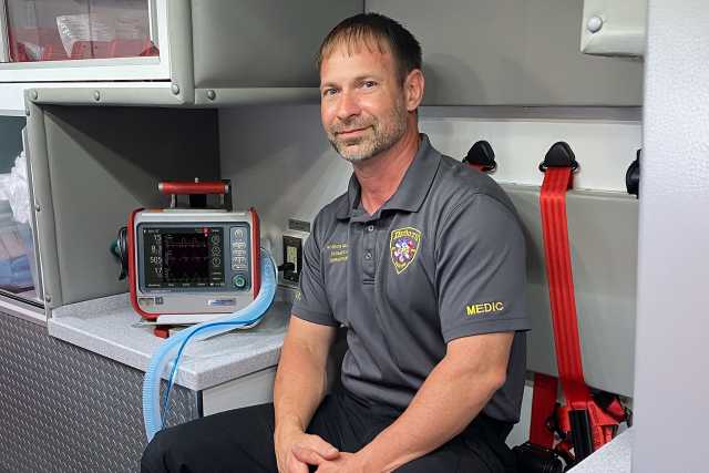
HAMILTON-T1.

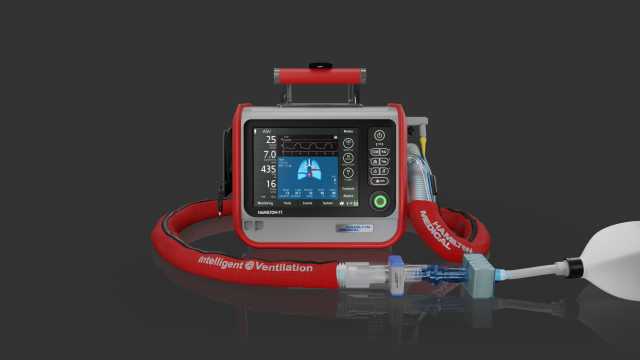
Ventilação segura no transporte de terapia intensiva

HAMILTON-T1

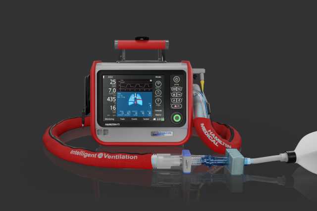
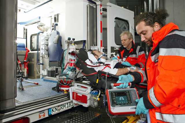
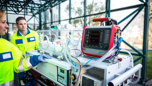
Nosso especialista em transporte! De neonatos a adultos

  • Respirador de UTI totalmente equipado para o transporte
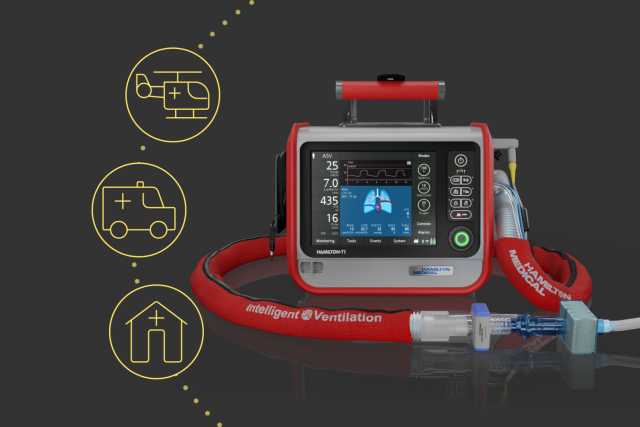
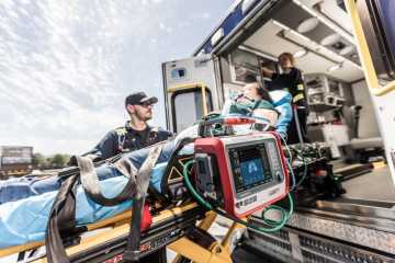
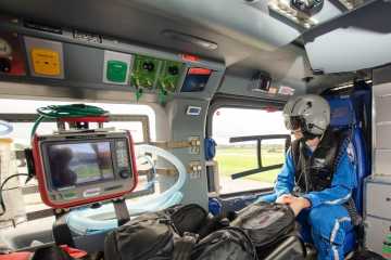
  • Aprovado para o transporte em terra, no ar e no mar
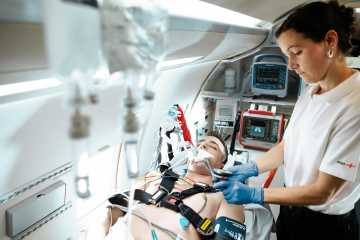
  • Dentro e fora do hospital
HAMILTON-T1
HAMILTON-T1

Nosso especialista em transporte! De neonatos a adultos

  • Respirador de UTI totalmente equipado para o transporte
  • Aprovado para o transporte do paciente em terra, no ar e no mar
  • Dentro e fora do hospital
HAMILTON-T1

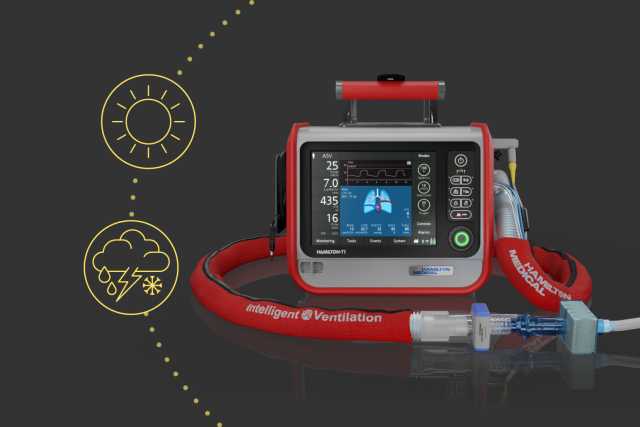
A seleção natural! Nas condições mais exigentes

  • Temperaturas de -15 °C a +50 °C
  • Proteção de entrada IP54
  • Altitude máxima de 7.620 metros
  • Invólucro reforçado e robusto com proteção contra impactos e vibrações
  • Resistente a choques, tela antirreflexo
HAMILTON-T1

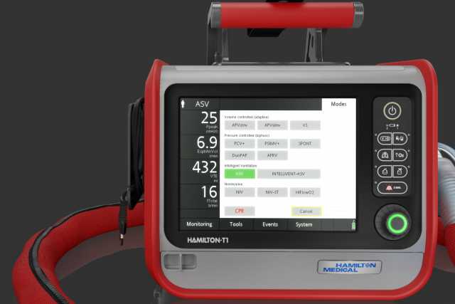
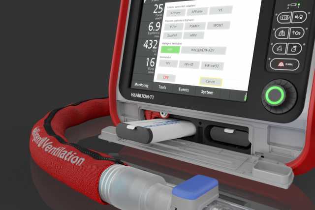
Tratamento de ventilação contínuo. Usa o mesmo modo e as mesmas configurações que junto do leito

  • Modos de ventilação de pressão controlada e com volume alvo
  • Ventilação adaptativa com ASV® e INTELLiVENT®-ASV
  • Ventilação não invasiva
  • Tratamento por cânula de alto fluxo nasal
HAMILTON-T1

Altamente independente. Sem ar comprimido e operado por bateria

  • Turbina de alto desempenho
  • Uma bateria integrada e uma bateria intercambiável a quente
  • Conector adicional para oxigênio de baixa pressão
HAMILTON-T1

A comunicação é a chave. Para uma melhor conexão

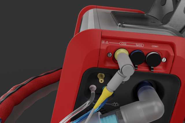
Opções de placa de comunicação para:

  • Sensores SpO2 e/ou CO2
  • Chamada à enfermagem
  • PDMS
  • HAMILTON-H900
  • RS232
HAMILTON-T1

Áreas de uso

A ter em conta

""

Pronto para a sua missão. Especificações à medida das suas necessidades

  • Dimensões (L x P x A):
    320 x 220 x 270 mm (unidade de ventilação)
    630 x 630 x 1380 mm (sem pega)
    630 x 630 x 1433 mm (com pega)

  • Peso:

    6,5 kg

  • Duração da bateria 4 h com uma bateria / 8 h com duas baterias

  • Opcional bateria intercambiável a quente

  • Todos os respiradores de transporte da Hamilton Medical estão equipados com o módulo Hamilton Connect.

  • Um protocolo de interface especialmente desenvolvido permite a conexão de dispositivos de terceiros para transferência de dados do respirador via Bluetooth.

  • Pressão controlada, volume alvo

  • Ventilação inteligente: ASV®, INTELLiVENT®‑ASV® (Não disponível em todos os mercadosA​), O2 assist (Não disponível em todos os mercadosA​)

  • Modos não invasivos (opcionalB​)

  • Tratamento de oxigênio de alto fluxo (opcionalB​)

  • Visualização da mecânica de pulmão (Pulmão Dinâmico)

  • Visualização da dependência do paciente em relação ao respirador

  • Capnografia volumétrica (opcionalB​)

  • Monitorização de SpO2 (opcionalB​)

Gostaria de obter mais informações?
Explore o modelo 3D

Descubra o HAMILTON-T1 de todos os ângulos e clique nos hotspots para saber mais.

Gráfico circular indicando que 71% das organizações de resgate aéreo (na Alemanha, Áustria, Suíça, Itália e Luxemburgo) escolheram o HAMILTON-T1 para seus helicópteros de terapia intensiva

A opção mais popular. Para 71% dos helicópteros de terapia intensiva

De acordo com o questionário HOVER (Handover of ventilated Helicopter Emergency Services [HEMS] — transferência de pacientes ventilados em serviços de emergência médica por helicópteros para a sala de emergênciaC ) realizado online entre organizações de resgate aéreo na Alemanha, Áustria, Suíça, Itália e Luxemburgo, 71% dessas organizações escolheram o HAMILTON-T1 para seus helicópteros de terapia intensiva (Hilbert-Carius, P., Struck, M.F., Hofer, V. et al. Nutzung des Hubschrauber-Respirators vom Landeplatz zum Zielort im Krankenhaus. Notfall Rettungsmed 23, 106–112 (2020). 1​).


Simplifique seu fluxo de trabalho com recursos inteligentes

Tratamento O2. Oxigenação no local

O tratamento O2 é uma ferramenta valiosa no ambiente pré-hospitalar, proporcionando melhor oxigenação para pacientes que necessitam, sem exigir intubação.

Essa opção permite realizar uma pré-oxigenação precisa de forma eficaz em campo.

Nebulizador pneumático. Para tratamentos adicionais

O fornecimento de uma névoa fina de partículas medicamentosas de aerossol pode ajudar a reverter o broncoespasmo, melhorar a eficiência da ventilação e reduzir a hipercapnia (Dhand R. New frontiers in aerosol delivery during mechanical ventilation. Respir Care. 2004;49(6):666-677. 100​​). 

Ventilação não invasiva. Sua primeira linha de defesa

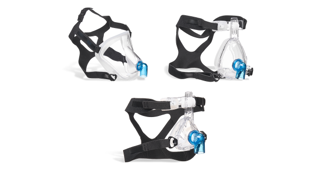
Escolha entre os modos VNI, VNI-ST (Somente disponível para HAMILTON-T1a​) ou CPAP (Somente disponível para HAMILTON-EM7b​) para fornecer suporte em diferentes cenários e garantir maior conforto ao paciente.

Em comparação com os respiradores que usam ar comprimido, os nossos respiradores acionados por turbina são capazes de fornecer taxas de fluxo de pico superior. Isto garante um desempenho intransigente, mesmo com fugas grandes.

Ventilação de RCP. Foque nas compressões, não no respirador

Foco, rapidez e precisão são essenciais em momentos críticos, enquanto as distrações devem ser minimizadas. A ventilação de RCP adapta os parâmetros de ventilação às situações em que a reanimação está sendo realizada, seguindo diretrizes internacionais (Del Rios M, Bartos JA, Panchal AR, Atkins DL, Cabanas JG, Cao D, Dainty KN, Dezfulian C, Donoghue AJ, Drennan IR, Elmer J, Hirsch KG, Idris AH, Joyner BL, Kamath-Rayne BD, Kleinman ME, Kurz MC, Lasa JJ, Lee HC, McBride ME, Raymond TT, Rittenberger, JC, Schexnayder SM, Szyld E, Topjian A, Wigginton JG, Previdi JK. Part 1: executive summary: 2025 American Heart Association Guidelines for Cardiopulmonary Resuscitation and Emergency Cardiovascular Care. Circulation. 2025;152(suppl):S284–S312. doi: 10.1161/CIR.0000000000001372 101​, Greif R, Lauridsen KG, Djärv T, et al. European Resuscitation Council Guidelines 2025 Executive Summary. Resuscitation. 2025;215 Suppl 1:110770. doi:10.1016/j.resuscitation.2025.110770102​).

Esta suporta você com acesso rápido e ajustes pré-configuráveis, ajuste adequado de alarme e disparo e exibição do timer RCP.

Capnografia baseada no tempo. Apoiando decisões mais seguras

A capnografia fornece monitorização contínua de etCO2 para avaliar a eficácia da ventilação, a integridade das vias aéreas e o estado do paciente em tempo real.

Oferece feedback imediato durante o suporte respiratório, a gestão de paragem cardíaca e o transporte, ajudando você a tomar decisões mais seguras e fundamentadas.

ASV — Adaptive Support Ventilation®. Suporte para você e seus pacientes

O ASV é um modo de ventilação adaptativo que ajusta continuamente os parâmetros de ventilação com base na mecânica de pulmão e no esforço do paciente.

Isso ajuda a simplificar a ventilação para equipes de transporte pré-hospitalar, oferecendo um único modo para pacientes intubados ativos e passivos, com apenas três parâmetros para controlar a eliminação de CO2 e a oxigenação.

O2 assist. Titulação de oxigênio mais inteligente na assistência pré-hospitalar

O O2 assist (Não disponível em todos os mercadosc​) é uma tecnologia avançada de gerenciamento de oxigênio desenvolvida para otimizar o fornecimento de oxigênio, inclusive durante o transporte.

Ao ajustar continuamente o fornecimento de oxigênio, o O2 assist mantém os níveis de SpO2 do paciente dentro de um intervalo-alvo definido. Isso reduz tanto o desperdício de oxigênio (Atakul G, Ceylan G, Sandal O, et al. Closed-loop oxygen usage during invasive mechanical ventilation of pediatric patients (CLOUDIMPP): a randomized controlled cross-over study. Front Med (Lausanne). 2024;11:1426969. Published 2024 Sep 10. doi:10.3389/fmed.2024.1426969103​, Trottier M, Bouchard PA, L'Her E, Lellouche F. Automated Oxygen Titration During CPAP and Noninvasive Ventilation in Healthy Subjects With Induced Hypoxemia. Respir Care. 2023;68(11):1553-1560. doi:10.4187/respcare.09866104​) quanto a necessidade de intervenções manuais (Roca O, Caritg O, Santafé M, et al. Closed-loop oxygen control improves oxygen therapy in acute hypoxemic respiratory failure patients under high flow nasal oxygen: a randomized cross-over study (the HILOOP study). Crit Care. 2022;26(1):108. Published 2022 Apr 14. doi:10.1186/s13054-022-03970-w105​).

Pulmão Dinâmico. Leitura imediata

O painel Pulmão Dinâmico traduz os dados de monitorização em uma representação visual de fácil interpretação.

Em um único gráfico, você pode visualizar complacência, resistência, SpO2, frequência de pulso e atividade espontânea do paciente.

Opção Óculos de visão noturna. Ver no escuro

Graças à opção de óculos de visão noturna (NVG), o respirador pode ser utilizado com segurança no escuro, sem afetar a visão do piloto nem interferir com os dispositivos de visão noturna.

Com o toque de um botão, o brilho da tela pode ser ajustado para um nível adequado ao uso com óculos de visão noturna.

INTELLiVENT®-ASV®. Seu assistente em movimento

Nosso modo de ventilação inteligente INTELLiVENT-ASV (Não disponível em todos os mercadosc​), vai mais além do ASV e permite-lhe passar de operador a supervisor.

Reduz o número de interações manuais com o respirador (Arnal JM, Garnero A, Novotni D, et al. Closed loop ventilation mode in Intensive Care Unit: a randomized controlled clinical trial comparing the numbers of manual ventilator setting changes. Minerva Anestesiol. 2018;84(1):58-67. doi:10.23736/S0375-9393.17.11963-2106​) e oferece ventilação para proteção pulmonar dos seus pacientes (Bialais E, Wittebole X, Vignaux L, et al. Closed-loop ventilation mode (IntelliVent®-ASV) in intensive care unit: a randomized trial. Minerva Anestesiol. 2016;82(6):657-668. 107​).

Oximetria de pulso. Para os entusiastas de SpO2

A opção SpO2 dispõe de medição não invasiva integrada de SpO2 com os dados exibidos de forma conveniente no seu respirador.
Permite detectar rapidamente a hipoxemia e avaliar a eficácia do tratamento de oxigênio durante o transporte.

Para pacientes com inalação de fumo, a medição de SpCO com a tecnologia rainbow da Masimo fornece valores não invasivos de carboxihemoglobina, auxiliando em decisões de triagem mais informadas.

Capnografia volumétrica. Elevando o cuidado ao paciente no ambiente pré-hospitalar

A capnografia volumétrica oferece uma visão contínua da ventilação e da perfusão ao medir o CO2 ao longo de toda a expiração, ajudando a identificar alterações na obstrução das vias aéreas, na eficiência da ventilação ou no estado de perfusão em tempo real (Blanch L, Romero PV, Lucangelo U. Volumetric capnography in the mechanically ventilated patient. Minerva Anestesiol. 2006;72(6):577-585. 108​).

Isso a torna especialmente valiosa durante o transporte de pacientes, onde as condições clínicas podem mudar rapidamente e a disponibilidade de dados imediatos e acionáveis é fundamental.

Alças e tendências configuráveis. Mantenha tudo sob controle

Na ventilação mecânica, alças e tendências fornecem informações em tempo real sobre o estado respiratório do paciente, permitindo identificar rapidamente problemas como obstrução das vias aéreas ou assincronia entre paciente–respirador.

Isso possibilita ajustes precisos da ventilação durante o transporte, contribuindo para melhores resultados e um atendimento mais seguro em ambientes pré-hospitalares dinâmicos.

Modos nCPAP. Suporte especializado para os mais pequenos

Com o modo nCPAP, seus pacientes mais pequenos recebem suporte com pressão positiva contínua nas vias aéreas. A adição do nCPAP ao seu respirador de transporte amplia as opções para o gerenciamento do suporte respiratório neonatal, de acordo com as melhores práticas atuais.

Gordon Miller

Customer voices

A disponibilidade dos modos não invasivos no HAMILTON-T1 me permitiu evitar muitas intubações, reduzir o risco para os pacientes e melhorar os resultados. Em 8 anos utilizando o HAMILTON-T1, precisei intubar apenas um paciente que era candidato à VNI no início do atendimento.

Gordon Miller

Supervisor and Training Office
DeSoto Parish EMS, Mansfield, LA, USA

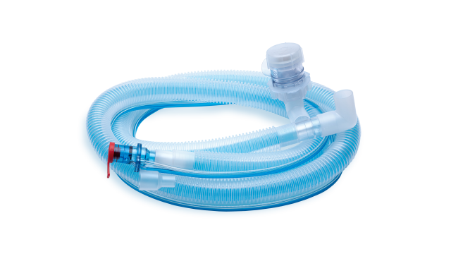
Consumíveis práticos

Consumíveis essenciais para operar o seu respirador

Os consumíveis essenciais da Hamilton Medical são concebidos para um desempenho ideal com os nossos respiradores. Para garantir a máxima satisfação do usuário e a segurança do paciente, projetamos os consumíveis para facilitar seu uso — de acordo com os mais elevados padrões de qualidade e segurança.

Iniciar seu treinamento sobre o respirador

""

Familiarize-se! Caminhos de aprendizagem e simulações

O Centro de aprendizagem do HAMILTON‑T1 oferece caminhos de aprendizagem fáceis de seguir para que possa conhecer o seu respirador e as suas tecnologias o mais rapidamente possível.

De seguida, experimente as suas novas competências em um ambiente seguro com o modelo de paciente virtual no nosso aplicativo VenTrainer!

Nossos serviços 360°

""

Prontos quando você estiver. Seu parceiro para cuidados de transporte pré-hospitalar

A equipe pré-hospitalar da Hamilton Medical está empenhada em fornecer serviços abrangentes e suporte para ventilação mecânica durante o transporte em contextos pré-hospitalares.

EMS_Prehospital-team_Contact-us

Tem alguma dúvida? Entre em contato!

Nossa equipe de atendimento pré-hospitalar está à disposição para responder às suas perguntas e oferecer consultoria especializada.

Footnotes

  • A. Não disponível em todos os mercados
  • B. Opcional para HAMILTON-T1
  • C. Transferência de pacientes ventilados em serviços de emergência médica por helicópteros para a sala de emergência
  • a. Somente disponível para HAMILTON-T1
  • b. Somente disponível para HAMILTON-EM7
  • c. Não disponível em todos os mercados

References

  1. 1. Hilbert-Carius, P., Struck, M.F., Hofer, V. et al. Nutzung des Hubschrauber-Respirators vom Landeplatz zum Zielort im Krankenhaus. Notfall Rettungsmed 23, 106–112 (2020).
  2. 100. Dhand R. New frontiers in aerosol delivery during mechanical ventilation. Respir Care. 2004;49(6):666-677.
  3. 101. Del Rios M, Bartos JA, Panchal AR, Atkins DL, Cabanas JG, Cao D, Dainty KN, Dezfulian C, Donoghue AJ, Drennan IR, Elmer J, Hirsch KG, Idris AH, Joyner BL, Kamath-Rayne BD, Kleinman ME, Kurz MC, Lasa JJ, Lee HC, McBride ME, Raymond TT, Rittenberger, JC, Schexnayder SM, Szyld E, Topjian A, Wigginton JG, Previdi JK. Part 1: executive summary: 2025 American Heart Association Guidelines for Cardiopulmonary Resuscitation and Emergency Cardiovascular Care. Circulation. 2025;152(suppl):S284–S312. doi: 10.1161/CIR.0000000000001372
  4. 102. Greif R, Lauridsen KG, Djärv T, et al. European Resuscitation Council Guidelines 2025 Executive Summary. Resuscitation. 2025;215 Suppl 1:110770. doi:10.1016/j.resuscitation.2025.110770
  5. 103. Atakul G, Ceylan G, Sandal O, et al. Closed-loop oxygen usage during invasive mechanical ventilation of pediatric patients (CLOUDIMPP): a randomized controlled cross-over study. Front Med (Lausanne). 2024;11:1426969. Published 2024 Sep 10. doi:10.3389/fmed.2024.1426969
  6. 104. Trottier M, Bouchard PA, L'Her E, Lellouche F. Automated Oxygen Titration During CPAP and Noninvasive Ventilation in Healthy Subjects With Induced Hypoxemia. Respir Care. 2023;68(11):1553-1560. doi:10.4187/respcare.09866
  7. 105. Roca O, Caritg O, Santafé M, et al. Closed-loop oxygen control improves oxygen therapy in acute hypoxemic respiratory failure patients under high flow nasal oxygen: a randomized cross-over study (the HILOOP study). Crit Care. 2022;26(1):108. Published 2022 Apr 14. doi:10.1186/s13054-022-03970-w
  8. 106. Arnal JM, Garnero A, Novotni D, et al. Closed loop ventilation mode in Intensive Care Unit: a randomized controlled clinical trial comparing the numbers of manual ventilator setting changes. Minerva Anestesiol. 2018;84(1):58-67. doi:10.23736/S0375-9393.17.11963-2
  9. 107. Bialais E, Wittebole X, Vignaux L, et al. Closed-loop ventilation mode (IntelliVent®-ASV) in intensive care unit: a randomized trial. Minerva Anestesiol. 2016;82(6):657-668.
  10. 108. Blanch L, Romero PV, Lucangelo U. Volumetric capnography in the mechanically ventilated patient. Minerva Anestesiol. 2006;72(6):577-585.

Nutzung des Hubschrauber-Respirators vom Landeplatz zum Zielort im Krankenhaus Sekundäranalyse der HOVER-Umfrage zu beatmeten Notfallpatienten in der Luftrettung

Hilbert-Carius, P., Struck, M.F., Hofer, V. et al. Nutzung des Hubschrauber-Respirators vom Landeplatz zum Zielort im Krankenhaus. Notfall Rettungsmed 23, 106–112 (2020).

New frontiers in aerosol delivery during mechanical ventilation.

Dhand R. New frontiers in aerosol delivery during mechanical ventilation. Respir Care. 2004;49(6):666-677.

The scientific basis for inhalation therapy in mechanically-ventilated patients is now firmly established. A variety of new devices that deliver drugs to the lung with high efficiency could be employed for drug delivery during mechanical ventilation. Encapsulation of drugs within liposomes could increase the amount of drug delivered, prolong the effect of a dose, and minimize adverse effects. With improved inhalation devices and surfactant formulations, inhaled surfactant could be employed for several indications in mechanically-ventilated patients. Research is unraveling the causes of some disorders that have been poorly understood, and our improved understanding of the causal mechanisms of various respiratory disorders will provide new applications for inhaled therapies.

2025 American Heart Association Guidelines for Cardiopulmonary Resuscitation and Emergency Cardiovascular Care

Del Rios M, Bartos JA, Panchal AR, Atkins DL, Cabanas JG, Cao D, Dainty KN, Dezfulian C, Donoghue AJ, Drennan IR, Elmer J, Hirsch KG, Idris AH, Joyner BL, Kamath-Rayne BD, Kleinman ME, Kurz MC, Lasa JJ, Lee HC, McBride ME, Raymond TT, Rittenberger, JC, Schexnayder SM, Szyld E, Topjian A, Wigginton JG, Previdi JK. Part 1: executive summary: 2025 American Heart Association Guidelines for Cardiopulmonary Resuscitation and Emergency Cardiovascular Care. Circulation. 2025;152(suppl):S284–S312. doi: 10.1161/CIR.0000000000001372

European Resuscitation Council Guidelines 2025 Executive Summary.

Greif R, Lauridsen KG, Djärv T, et al. European Resuscitation Council Guidelines 2025 Executive Summary. Resuscitation. 2025;215 Suppl 1:110770. doi:10.1016/j.resuscitation.2025.110770

The 2025 European Resuscitation Council (ERC) Guidelines present the most up-to-date evidence-based guidelines for the practice of resuscitation across Europe. The ERC Guidelines 2025 are based on evidence produced by the International Liaison Committee on Resuscitation (ILCOR) in the form of systematic reviews, scoping reviews, and evidence updates, published as the ILCOR Consensus on Science with Treatment Recommendations. The certainty of evidence of these ILCOR treatment recommendations was used to issue the ERC Guidelines 2025 Recommendations. In some cases, the ERC made good practice statements when evidence was absent for certain topics. If no ILCOR review was available, the ERC writing groups conducted their own reviews to provide recommendations. The ERC Guidelines 2025 cover the epidemiology of cardiac arrest, the role that systems play in saving lives, adult basic life support, adult advanced life support, resuscitation in special circumstances, post resuscitation care, newborn resuscitation and support of transition of infants at birth, paediatric basic and advanced life support, resuscitation ethics, education for resuscitation, and first aid. These guidelines are a framework of recommendations for the approach to out-of-hospital and in-hospital resuscitation; the implementation is achieved locally taking local legislation and health care regulations into consideration.

Closed-loop oxygen usage during invasive mechanical ventilation of pediatric patients (CLOUDIMPP): a randomized controlled cross-over study.

Atakul G, Ceylan G, Sandal O, et al. Closed-loop oxygen usage during invasive mechanical ventilation of pediatric patients (CLOUDIMPP): a randomized controlled cross-over study. Front Med (Lausanne). 2024;11:1426969. Published 2024 Sep 10. doi:10.3389/fmed.2024.1426969

BACKGROUND The aim of this study is the evaluation of a closed-loop oxygen control system in pediatric patients undergoing invasive mechanical ventilation (IMV). METHODS Cross-over, multicenter, randomized, single-blind clinical trial. Patients between the ages of 1 month and 18 years who were undergoing IMV therapy for acute hypoxemic respiratory failure (AHRF) were assigned at random to either begin with a 2-hour period of closed-loop oxygen control or manual oxygen titrations. By using closed-loop oxygen control, the patients' SpO2 levels were maintained within a predetermined target range by the automated adjustment of the FiO2. During the manual oxygen titration phase of the trial, healthcare professionals at the bedside made manual changes to the FiO2, while maintaining the same target range for SpO2. Following either period, the patient transitioned to the alternative therapy. The outcomes were the percentage of time spent in predefined SpO2 ranges ±2% (primary), FiO2, total oxygen use, and the number of manual adjustments. FINDINGS The median age of included 33 patients was 17 (13-55.5) months. In contrast to manual oxygen titrations, patients spent a greater proportion of time within a predefined optimal SpO2 range when the closed-loop oxygen controller was enabled (95.7% [IQR 92.1-100%] vs. 65.6% [IQR 41.6-82.5%]), mean difference 33.4% [95%-CI 24.5-42%]; P < 0.001). Median FiO2 was lower (32.1% [IQR 23.9-54.1%] vs. 40.6% [IQR 31.1-62.8%]; P < 0.001) similar to total oxygen use (19.8 L/h [IQR 4.6-64.8] vs. 39.4 L/h [IQR 16.8-79]; P < 0.001); however, median SpO2/FiO2 was higher (329.4 [IQR 180-411.1] vs. 246.7 [IQR 151.1-320.5]; P < 0.001) with closed-loop oxygen control. With closed-loop oxygen control, the median number of manual adjustments reduced (0.0 [IQR 0.0-0.0] vs. 1 [IQR 0.0-2.2]; P < 0.001). CONCLUSION Closed-loop oxygen control enhances oxygen therapy in pediatric patients undergoing IMV for AHRF, potentially leading to more efficient utilization of oxygen. This technology also decreases the necessity for manual adjustments, which could reduce the workloads of healthcare providers. CLINICAL TRIAL REGISTRATION This research has been submitted to ClinicalTrials.gov (NCT05714527).

Automated Oxygen Titration During CPAP and Noninvasive Ventilation in Healthy Subjects With Induced Hypoxemia.

Trottier M, Bouchard PA, L'Her E, Lellouche F. Automated Oxygen Titration During CPAP and Noninvasive Ventilation in Healthy Subjects With Induced Hypoxemia. Respir Care. 2023;68(11):1553-1560. doi:10.4187/respcare.09866

BACKGROUND Automated oxygen titration to maintain a stable SpO2 has been developed for spontaneously breathing patients but has not been evaluated during CPAP and noninvasive ventilation (NIV). METHODS We performed a randomized controlled crossover, double-blind study on 10 healthy subjects with induced hypoxemia during 3 situations: spontaneous breathing with oxygen support, CPAP (5 cm H2O), and NIV (7/3 cm H2O). We conducted in random order 3 dynamic hypoxic challenges of 5 min (FIO2 0.08 ± 0.02, 0.11± 0.02, and 0.14 ± 0.02). For each condition, we compared automated oxygen titration and manual oxygen titration by experienced respiratory therapists (RTs), with the aim to maintain the SpO2 at 94 ± 2%. In addition, we included 2 subjects hospitalized for exacerbation of COPD under NIV and a subject managed after bariatric surgery with CPAP and automated oxygen titration. RESULTS The percentage of time in the SpO2 target was higher with automated compared with manual oxygen titration for all conditions, on average 59.6 ± 22.8% compared to 44.3 ± 23.9% (P = .004). Hyperoxemia (SpO2 > 96%) was less frequent with automated titration for each mode of oxygen administration (24.0 ± 24.4% vs 39.1 ± 25.3%, P < .001). During the manual titration periods, the RT made several changes to oxygen flow (5.1 ± 3.3 interventions that lasted 122 ± 70 s/period) compared to none during the automated titration to maintain oxygenation in the targeted SpO2 . Time in the SpO2 target was higher with stable hospitalized subjects in comparison with healthy subjects under dynamic-induced hypoxemia. CONCLUSIONS In this proof-of-concept study, automated oxygen titration was used during CPAP and NIV. The performances to maintain the SpO2 target were significantly better compared to manual oxygen titration in the setting of this study protocol. This technology may allow decreasing the number of manual interventions for oxygen titration during CPAP and NIV.

Closed-loop oxygen control improves oxygen therapy in acute hypoxemic respiratory failure patients under high flow nasal oxygen: a randomized cross-over study (the HILOOP study).

Roca O, Caritg O, Santafé M, et al. Closed-loop oxygen control improves oxygen therapy in acute hypoxemic respiratory failure patients under high flow nasal oxygen: a randomized cross-over study (the HILOOP study). Crit Care. 2022;26(1):108. Published 2022 Apr 14. doi:10.1186/s13054-022-03970-w

BACKGROUND We aimed to assess the efficacy of a closed-loop oxygen control in critically ill patients with moderate to severe acute hypoxemic respiratory failure (AHRF) treated with high flow nasal oxygen (HFNO). METHODS In this single-centre, single-blinded, randomized crossover study, adult patients with moderate to severe AHRF who were treated with HFNO (flow rate ≥ 40 L/min with FiO2 ≥ 0.30) were randomly assigned to start with a 4-h period of closed-loop oxygen control or 4-h period of manual oxygen titration, after which each patient was switched to the alternate therapy. The primary outcome was the percentage of time spent in the individualized optimal SpO2 range. RESULTS Forty-five patients were included. Patients spent more time in the optimal SpO2 range with closed-loop oxygen control compared with manual titrations of oxygen (96.5 [93.5 to 98.9] % vs. 89 [77.4 to 95.9] %; p < 0.0001) (difference estimate, 10.4 (95% confidence interval 5.2 to 17.2). Patients spent less time in the suboptimal range during closed-loop oxygen control, both above and below the cut-offs of the optimal SpO2 range, and less time above the suboptimal range. Fewer number of manual adjustments per hour were needed with closed-loop oxygen control. The number of events of SpO2 < 88% and < 85% were not significantly different between groups. CONCLUSIONS Closed-loop oxygen control improves oxygen administration in patients with moderate-to-severe AHRF treated with HFNO, increasing the percentage of time in the optimal oxygenation range and decreasing the workload of healthcare personnel. These results are especially relevant in a context of limited oxygen supply and high medical demand, such as the COVID-19 pandemic. Trial registration The HILOOP study was registered at www. CLINICALTRIALS gov under the identifier NCT04965844 .

Closed loop ventilation mode in Intensive Care Unit: a randomized controlled clinical trial comparing the numbers of manual ventilator setting changes.

Arnal JM, Garnero A, Novotni D, et al. Closed loop ventilation mode in Intensive Care Unit: a randomized controlled clinical trial comparing the numbers of manual ventilator setting changes. Minerva Anestesiol. 2018;84(1):58-67. doi:10.23736/S0375-9393.17.11963-2

BACKGROUND There is an equipoise regarding closed-loop ventilation modes and the ability to reduce workload for providers. On one hand some settings are managed by the ventilator but on another hand the automatic mode introduces new settings for the user. METHODS This randomized controlled trial compared the number of manual ventilator setting changes between a full closed loop ventilation and oxygenation mode (INTELLiVENT-ASV®) and conventional ventilation modes (volume assist control and pressure support) in Intensive Care Unit (ICU) patients. The secondary endpoints were to compare the number of arterial blood gas analysis, the sedation dose and the user acceptance. Sixty subjects with an expected duration of mechanical ventilation of at least 48 hours were randomized to be ventilated using INTELLiVENT-ASV® or conventional modes with a protocolized weaning. All manual ventilator setting changes were recorded continuously from inclusion to successful extubation or death. Arterial blood gases were performed upon decision of the clinician in charge. User acceptance score was assessed for nurses and physicians once daily using a Likert Scale. RESULTS The number of manual ventilator setting changes per 24 h-period per subject was lower in INTELLiVENT-ASV® as compared to conventional ventilation group (5 [4-7] versus 10 [7-17]) manuals settings per subject per day [P<0.001]). The number of arterial blood gas analysis and the sedation doses were not significantly different between the groups. Nurses and physicians reported that INTELLiVENT-ASV® was significantly easier to use as compared to conventional ventilation (P<0.001 for nurses and P<0.01 for physicians). CONCLUSIONS For mechanically ventilated ICU patients, INTELLiVENT-ASV® significantly reduces the number of manual ventilator setting changes with the same number of arterial blood gas analysis and sedation dose, and is easier to use for the caregivers as compared to conventional ventilation modes.

Closed-loop ventilation mode (IntelliVent®-ASV) in intensive care unit: a randomized trial.

Bialais E, Wittebole X, Vignaux L, et al. Closed-loop ventilation mode (IntelliVent®-ASV) in intensive care unit: a randomized trial. Minerva Anestesiol. 2016;82(6):657-668.

BACKGROUND Closed-loop modes automatically adjust ventilation settings, delivering individualized ventilation over short periods of time. The objective of this randomized controlled trial was to compare safety, efficacy and workload for the health care team between IntelliVent®-ASV and conventional modes over a 48-hour period. METHODS ICU patients admitted with an expected duration of mechanical ventilation of more than 48 hours were randomized to IntelliVent®-ASV or conventional ventilation modes. All ventilation parameters were recorded breath-by-breath. The number of manual adjustments assesses workload for the healthcare team. Safety and efficacy were assessed by calculating the time spent within previously defined ranges of non-optimal and optimal ventilation, respectively. RESULTS Eighty patients were analyzed. The median values of ventilation parameters over 48 hours were similar in both groups except for PEEP (7[4] cmH2O versus 6[3] cmH2O with IntelliVent®-ASV and conventional ventilation, respectively, P=0.028) and PETCO2 (36±7 mmHg with IntelliVent®-ASV versus 40±8 mmHg with conventional ventilation, P=0.041). Safety was similar between IntelliVent®-ASV and conventional ventilation for all parameters except for PMAX, which was more often non-optimal with IntelliVent®-ASV (P=0.001). Efficacy was comparable between the 2 ventilation strategies, except for SpO2 and VT, which were more often optimal with IntelliVent®-ASV (P=0.005, P=0.016, respectively). IntelliVent®-ASV required less manual adjustments than conventional ventilation (P<0.001) for a higher total number of adjustments (P<0.001). The coefficient of variation over 48 hours was larger with IntelliVent®-ASV in regard of maximum pressure, inspiratory pressure (PINSP), and PEEP as compared to conventional ventilation. CONCLUSIONS IntelliVent®-ASV required less manual intervention and delivered more variable PEEP and PINSP, while delivering ventilation safe and effective ventilation in terms of VT, RR, SpO2 and PETCO2.

Volumetric capnography in the mechanically ventilated patient.

Blanch L, Romero PV, Lucangelo U. Volumetric capnography in the mechanically ventilated patient. Minerva Anestesiol. 2006;72(6):577-585.

Expiratory capnogram provides qualitative information on the waveform patterns associated with mechanical ventilation and quantitative estimation of expired CO2. Volumetric capnography simultaneously measures expired CO2 and tidal volume and allows identification of CO2 from 3 sequential lung compartments: apparatus and anatomic dead space, from progressive emptying of alveoli and alveolar gas. Lung heterogeneity creates regional differences in CO2 concentration and sequential emptying contributes to the rise of the alveolar plateau and to the steeper the expired CO2 slope. The concept of dead space accounts for those lung areas that are ventilated but not perfused. In patients with sudden pulmonary vascular occlusion due to pulmonary embolism, the resultant high V/Q mismatch produces an increase in alveolar dead space. Calculations derived from volumetric capnography are useful to suspect pulmonary embolism at the bedside. Alveolar dead space is large in acute lung injury and when the effect of positive end-expiratory pressure (PEEP) is to recruit collapsed lung units resulting in an improvement of oxygenation, alveolar dead space may decrease, whereas PEEP-induced overdistension tends to increase alveolar dead space. Finally, measurement of physiologic dead space and alveolar ejection volume at admission or the trend during the first 48 hours of mechanical ventilation might provide useful information on outcome of critically ill patients with acute lung injury or acute respiratory distress syndrome.