Мы обнаружили, что вы посещаете наш сайт из страны Соединенные Штаты.
Для вашей страны (Соединенные Штаты) доступна отдельная версия веб-сайта.

 Технологии

Транспульмональное давление. Подробное объяснение механики дыхательной системы

Катетер NutriVent для измерения пищеводного давления

Взгляд изнутри. Измерение пищеводного давления

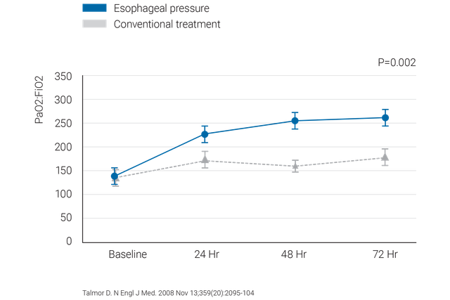
Измерение пищеводного давления (Pes) — это малоинвазивный метод мониторинга, который позволяет определить транспульмональное давление.

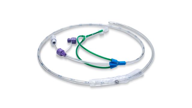
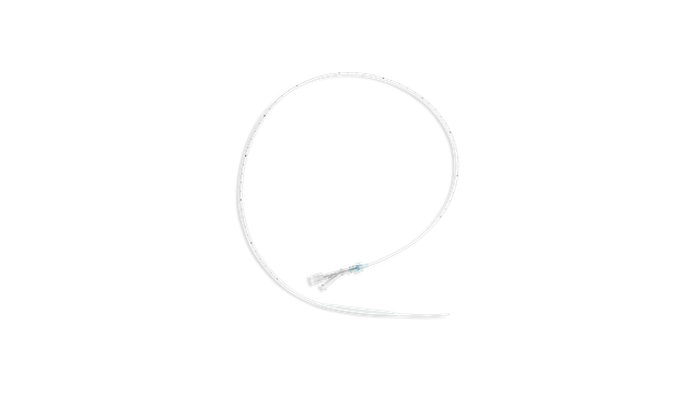
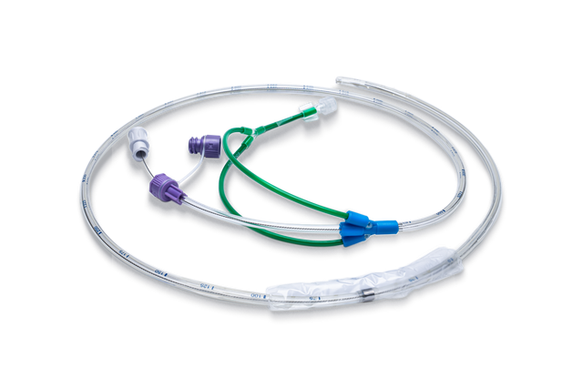
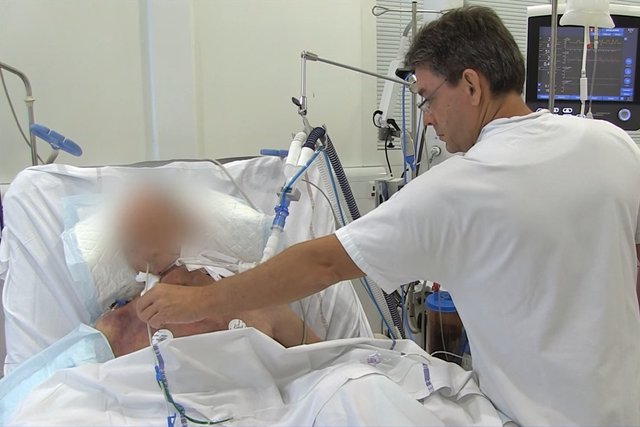
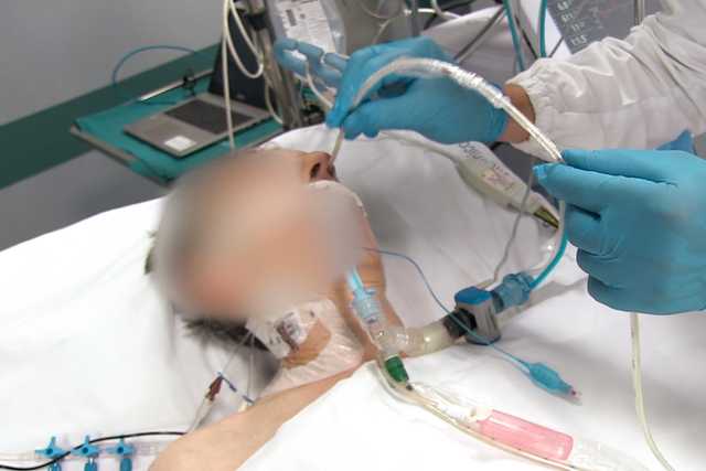
Наиболее распространенный способ измерения значения Pes — с помощью наполненного воздухом баллона, встроенного в пищеводный катетер.

Экран аппарата ИВЛ, на котором в виде кривой отображены пищеводное давление (Pes) и транспульмональное давление (Ptranspulm). Экран аппарата ИВЛ, на котором в виде кривой отображены пищеводное давление (Pes) и транспульмональное давление (Ptranspulm).

Лучше один раз увидеть, чем сто раз услышать! Значения Pes и Ptranspulm на экране

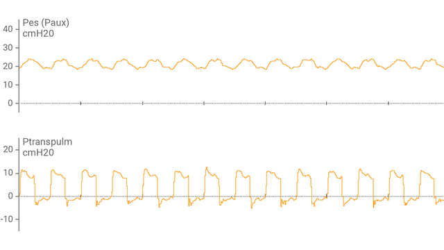
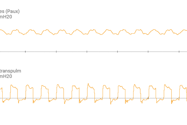
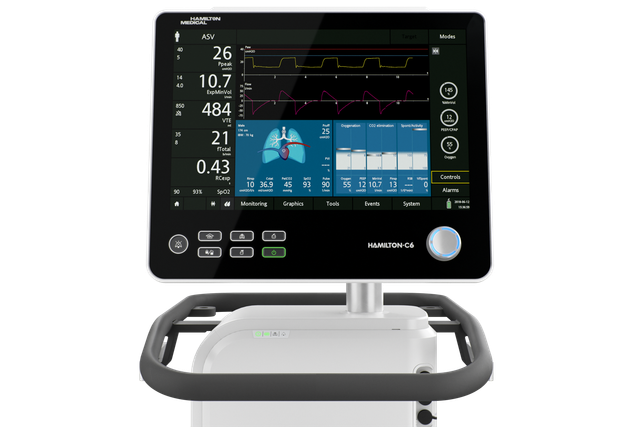
После подсоединения пищеводного баллонного катетера к вспомогательному порту и подтверждения правильности размещения на экране аппарата ИВЛ в виде кривой отобразится пищеводное давление (Pes) и транспульмональное давление (Ptranspulm).

Измерить статическое значение Ptranspulm можно с помощью маневров задержки вдоха и выдоха.

Врач и пациент

Консолидация усилий! Оценка возможности раскрытия объема легких

Для оценки возможности раскрытия объема легких и выполнения маневров рекрутмента также можно использовать транспульмональное давление в сочетании с инструментом P/V Tool® Pro.

Графическое представление статистических данных: Talmor D. N Engl J Med. 2008 Nov 13;359(20):2095-104

Каковы преимущества? Немного доказательств

  • В результате настройки параметра PEEP на основе транспульмонального давления улучшились податливость и оксигенация у пациентов с ARDS (Talmor D, Sarge T, Malhotra A, et al. Mechanical ventilation guided by esophageal pressure in acute lung injury. N Engl J Med. 2008;359(20):2095-2104. doi:10.1056/NEJMoa07086381​).
  • После установки целевого значения транспульмонального давления улучшились эластичность и рабочее давление. Также можно сделать вывод о снижении уровня смертности за период 28 дней (Baedorf Kassis E, Loring SH, Talmor D. Mortality and pulmonary mechanics in relation to respiratory system and transpulmonary driving pressures in ARDS. Intensive Care Med. 2016;42(8):1206-1213. doi:10.1007/s00134-016-4403-72​).
  • Мониторинг транспульмонального давления может помочь избежать использования экстракорпоральной мембранной оксигенации (ЭКМО) у пациентов в очень тяжелом состоянии (Grasso S, Terragni P, Birocco A, et al. ECMO criteria for influenza A (H1N1)-associated ARDS: role of transpulmonary pressure. Intensive Care Med. 2012;38(3):395-403. doi:10.1007/s00134-012-2490-73​).
  • Стратегия вентиляции с управлением по транспульмональному давлению может увеличить долю пациентов с тяжелым острым респираторным дистресс-синдромом (ARDS), успешно отлученных от ЭКМО (Wang R, Sun B, Li X, et al. Mechanical Ventilation Strategy Guided by Transpulmonary Pressure in Severe Acute Respiratory Distress Syndrome Treated With Venovenous Extracorporeal Membrane Oxygenation. Crit Care Med. 2020;48(9):1280-1288. doi:10.1097/CCM.00000000000044454​).
  • Измерение пищеводного давления (Pes) – это золотой стандарт оценки дыхательных усилий и работы дыхательной системы (Bertoni M, Spadaro S, Goligher EC. Monitoring Patient Respiratory Effort During Mechanical Ventilation: Lung and Diaphragm-Protective Ventilation. Crit Care. 2020;24(1):106. Published 2020 Mar 24. doi:10.1186/s13054-020-2777-y5​).
Изображение студента с сертификатом в руке

Полезно знать! Материалы курса обучения измерению транспульмонального давления

Watch this short demonstration to learn how to use transpulmonary pressure measurement to set PEEP in mechanically ventilated patients more accurately.

Установка значения PEEP на основе транспульмонального давления

Посмотрите этот короткий ролик, чтобы узнать, как использовать мониторинг транспульмонального давления для более точной установки значения PEEP.

Установка пищеводного баллонного катетера в пищевод пациента

Это вопрос техники! Установка пищеводного баллонного катетера

Электронная книга, которая содержит 13 советов от специалистов

Бесплатная электронная книга

13 советов от специалистов. Измерение пищеводного давления

Клинически проверенные рекомендации касательно того, что следует делать и чего следует избегать при измерении пищеводного давления у пациентов с острым респираторным дистресс-синдромом.

Расходные материалы

Компания Hamilton Medical предлагает пищеводные баллонные катетеры производства компании Cooper Surgical и назогастральные катетеры производства компании NutriVent.

Доступность

Мониторинг транспульмонального давления – это стандартная функция в аппаратах ИВЛ HAMILTON-C6 и HAMILTON-G5/S1.

Mechanical ventilation guided by esophageal pressure in acute lung injury.

Talmor D, Sarge T, Malhotra A, et al. Mechanical ventilation guided by esophageal pressure in acute lung injury. N Engl J Med. 2008;359(20):2095-2104. doi:10.1056/NEJMoa0708638


BACKGROUND

Survival of patients with acute lung injury or the acute respiratory distress syndrome (ARDS) has been improved by ventilation with small tidal volumes and the use of positive end-expiratory pressure (PEEP); however, the optimal level of PEEP has been difficult to determine. In this pilot study, we estimated transpulmonary pressure with the use of esophageal balloon catheters. We reasoned that the use of pleural-pressure measurements, despite the technical limitations to the accuracy of such measurements, would enable us to find a PEEP value that could maintain oxygenation while preventing lung injury due to repeated alveolar collapse or overdistention.

METHODS

We randomly assigned patients with acute lung injury or ARDS to undergo mechanical ventilation with PEEP adjusted according to measurements of esophageal pressure (the esophageal-pressure-guided group) or according to the Acute Respiratory Distress Syndrome Network standard-of-care recommendations (the control group). The primary end point was improvement in oxygenation. The secondary end points included respiratory-system compliance and patient outcomes.

RESULTS

The study reached its stopping criterion and was terminated after 61 patients had been enrolled. The ratio of the partial pressure of arterial oxygen to the fraction of inspired oxygen at 72 hours was 88 mm Hg higher in the esophageal-pressure-guided group than in the control group (95% confidence interval, 78.1 to 98.3; P=0.002). This effect was persistent over the entire follow-up time (at 24, 48, and 72 hours; P=0.001 by repeated-measures analysis of variance). Respiratory-system compliance was also significantly better at 24, 48, and 72 hours in the esophageal-pressure-guided group (P=0.01 by repeated-measures analysis of variance).

CONCLUSIONS

As compared with the current standard of care, a ventilator strategy using esophageal pressures to estimate the transpulmonary pressure significantly improves oxygenation and compliance. Multicenter clinical trials are needed to determine whether this approach should be widely adopted. (ClinicalTrials.gov number, NCT00127491.)

Mortality and pulmonary mechanics in relation to respiratory system and transpulmonary driving pressures in ARDS.

Baedorf Kassis E, Loring SH, Talmor D. Mortality and pulmonary mechanics in relation to respiratory system and transpulmonary driving pressures in ARDS. Intensive Care Med. 2016;42(8):1206-1213. doi:10.1007/s00134-016-4403-7


PURPOSE

The driving pressure of the respiratory system has been shown to strongly correlate with mortality in a recent large retrospective ARDSnet study. Respiratory system driving pressure [plateau pressure-positive end-expiratory pressure (PEEP)] does not account for variable chest wall compliance. Esophageal manometry can be utilized to determine transpulmonary driving pressure. We have examined the relationships between respiratory system and transpulmonary driving pressure, pulmonary mechanics and 28-day mortality.

METHODS

Fifty-six patients from a previous study were analyzed to compare PEEP titration to maintain positive transpulmonary end-expiratory pressure to a control protocol. Respiratory system and transpulmonary driving pressures and pulmonary mechanics were examined at baseline, 5 min and 24 h. Analysis of variance and linear regression were used to compare 28 day survivors versus non-survivors and the intervention group versus the control group, respectively.

RESULTS

At baseline and 5 min there was no difference in respiratory system or transpulmonary driving pressure. By 24 h, survivors had lower respiratory system and transpulmonary driving pressures. Similarly, by 24 h the intervention group had lower transpulmonary driving pressure. This decrease was explained by improved elastance and increased PEEP.

CONCLUSIONS

The results suggest that utilizing PEEP titration to target positive transpulmonary pressure via esophageal manometry causes both improved elastance and driving pressures. Treatment strategies leading to decreased respiratory system and transpulmonary driving pressure at 24 h may be associated with improved 28 day mortality. Studies to clarify the role of respiratory system and transpulmonary driving pressures as a prognosticator and bedside ventilator target are warranted.

ECMO criteria for influenza A (H1N1)-associated ARDS: role of transpulmonary pressure.

Grasso S, Terragni P, Birocco A, et al. ECMO criteria for influenza A (H1N1)-associated ARDS: role of transpulmonary pressure. Intensive Care Med. 2012;38(3):395-403. doi:10.1007/s00134-012-2490-7


PURPOSE

To assess whether partitioning the elastance of the respiratory system (E (RS)) between lung (E (L)) and chest wall (E (CW)) elastance in order to target values of end-inspiratory transpulmonary pressure (PPLAT(L)) close to its upper physiological limit (25 cmH(2)O) may optimize oxygenation allowing conventional treatment in patients with influenza A (H1N1)-associated ARDS referred for extracorporeal membrane oxygenation (ECMO).

METHODS

Prospective data collection of patients with influenza A (H1N1)-associated ARDS referred for ECMO (October 2009-January 2010). Esophageal pressure was used to (a) partition respiratory mechanics between lung and chest wall, (b) titrate positive end-expiratory pressure (PEEP) to target the upper physiological limit of PPLAT(L) (25 cmH(2)O).

RESULTS

Fourteen patients were referred for ECMO. In seven patients PPLAT(L) was 27.2 ± 1.2 cmH(2)O; all these patients underwent ECMO. In the other seven patients, PPLAT(L) was 16.6 ± 2.9 cmH(2)O. Raising PEEP (from 17.9 ± 1.2 to 22.3 ± 1.4 cmH(2)O, P = 0.0001) to approach the upper physiological limit of transpulmonary pressure (PPLAT(L) = 25.3 ± 1.7 cm H(2)O) improved oxygenation index (from 37.4 ± 3.7 to 16.5 ± 1.4, P = 0.0001) allowing patients to be treated with conventional ventilation.

CONCLUSIONS

Abnormalities of chest wall mechanics may be present in some patients with influenza A (H1N1)-associated ARDS. These abnormalities may not be inferred from measurements of end-inspiratory plateau pressure of the respiratory system (PPLAT(RS)). In these patients, titrating PEEP to PPLAT(RS) may overestimate the incidence of hypoxemia refractory to conventional ventilation leading to inappropriate use of ECMO.

Mechanical Ventilation Strategy Guided by Transpulmonary Pressure in Severe Acute Respiratory Distress Syndrome Treated With Venovenous Extracorporeal Membrane Oxygenation.

Wang R, Sun B, Li X, et al. Mechanical Ventilation Strategy Guided by Transpulmonary Pressure in Severe Acute Respiratory Distress Syndrome Treated With Venovenous Extracorporeal Membrane Oxygenation. Crit Care Med. 2020;48(9):1280-1288. doi:10.1097/CCM.0000000000004445


OBJECTIVES

Previous studies have suggested that adjusting ventilator settings based on transpulmonary pressure measurements may minimize ventilator-induced lung injury, but this has never been investigated in patients with severe acute respiratory distress syndrome supported with venovenous extracorporeal membrane oxygenation. We aimed to evaluate whether a transpulmonary pressure-guided ventilation strategy would increase the proportion of patients successfully weaned from venovenous extracorporeal membrane oxygenation support in patients with severe acute respiratory distress syndrome.

DESIGN

Single-center, prospective, randomized controlled trial.

SETTING

Sixteen-bed, respiratory ICU at a tertiary academic medical center.

PATIENTS

Severe acute respiratory distress syndrome patients receiving venovenous extracorporeal membrane oxygenation.

INTERVENTIONS

One-hundred four patients were randomized to transpulmonary pressure-guided ventilation group (n = 52) or lung rest strategy group (n = 52) groups. Two patients had cardiac arrest during establishment of venovenous extracorporeal membrane oxygenation in the lung rest group did not receive the assigned intervention. Thus, 102 patients were included in the analysis.

MEASUREMENTS AND MAIN RESULTS

The proportion of patients successfully weaned from venovenous extracorporeal membrane oxygenation in the transpulmonary pressure-guided group was significantly higher than that in the lung rest group (71.2% vs 48.0%; p = 0.017). Compared with the lung rest group, driving pressure, tidal volumes, and mechanical power were significantly lower, and positive end-expiratory pressure was significantly higher, in the transpulmonary pressure-guided group during venovenous extracorporeal membrane oxygenation support. In the transpulmonary pressure-guided group, levels of interleukin-1β, interleukin-6, and interleukin-8 were significantly lower, and interleukin-10 was significantly higher, than those of the lung rest group over time. Lung density was significantly lower in the transpulmonary pressure-guided group after venovenous extracorporeal membrane oxygenation support than in the lung rest group.

CONCLUSIONS

A transpulmonary pressure-guided ventilation strategy could increase the proportion of patients with severe acute respiratory distress syndrome successfully weaned from venovenous extracorporeal membrane oxygenation.

Monitoring Patient Respiratory Effort During Mechanical Ventilation: Lung and Diaphragm-Protective Ventilation.

Bertoni M, Spadaro S, Goligher EC. Monitoring Patient Respiratory Effort During Mechanical Ventilation: Lung and Diaphragm-Protective Ventilation. Crit Care. 2020;24(1):106. Published 2020 Mar 24. doi:10.1186/s13054-020-2777-y

This article is one of ten reviews selected from the Annual Update in Intensive Care and Emergency Medicine 2020. Other selected articles can be found online at https://www.biomedcentral.com/collections/annualupdate2020. Further information about the Annual Update in Intensive Care and Emergency Medicine is available from http://www.springer.com/series/8901.